What are the cervical spinal joints?
Generally, there are three cervical spinal joints located between each two adjacent vertebrae: one disc joint (intervertebral disc joint) and two paired (left and right) facet joints. The disc joint is located anteriorly and the facet joints are located posterolaterally (Fig. 5).
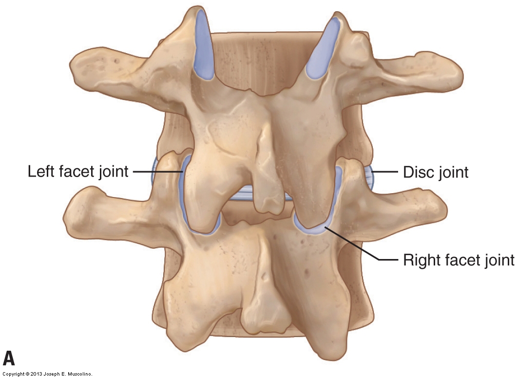
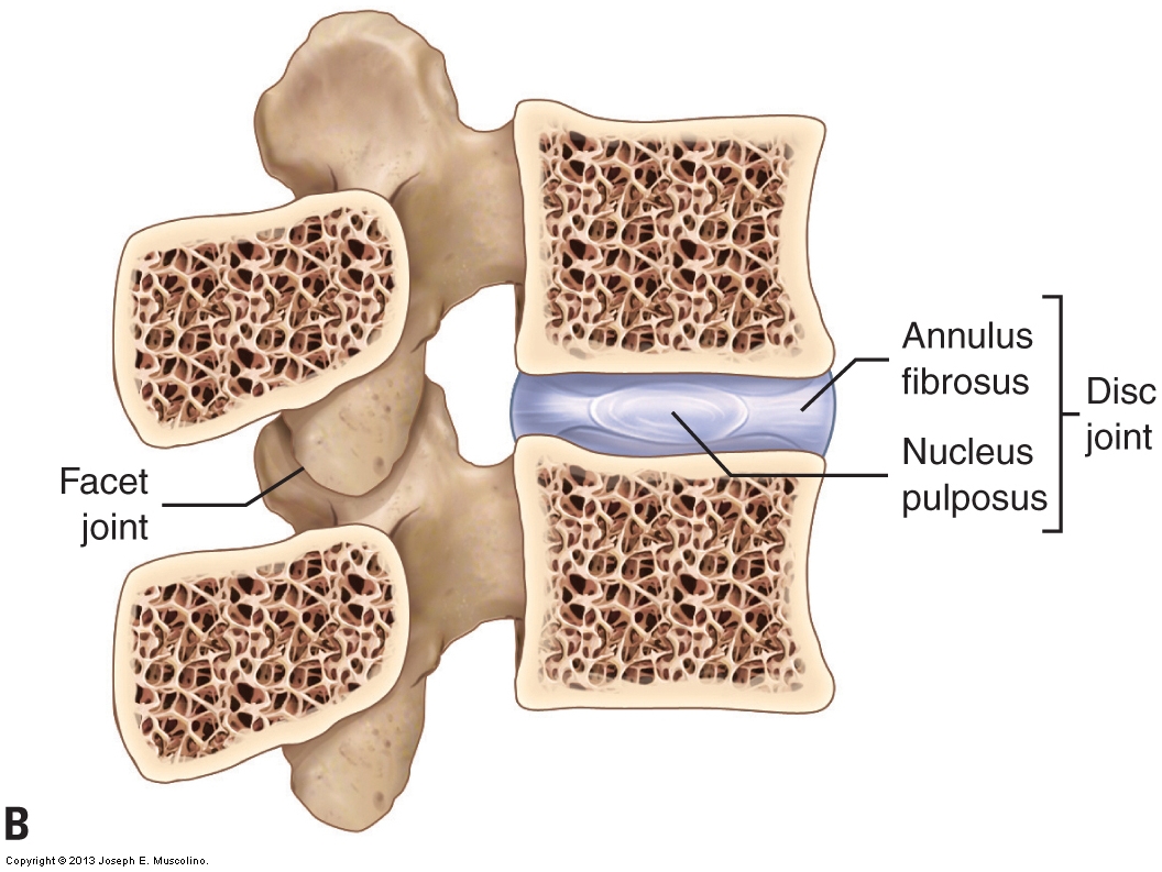
Figure 5. The disc and facet joints of the spine. A is a posterior view. The disc joint is located anteriorly; the paired facet joints are located posterolaterally. B is a right lateral view of a sagittal plane cross section. The disc joint is composed of the outer annulus fibrosus fibers and an inner nucleus pulposus. (Courtesy of Joseph E. Muscolino – Manual Therapy for the Low Back and Pelvis – A Clinical Orthopedic Approach 2015.)
Cervical Spinal Joints – Disc Joint
The disc joint is a cartilaginous joint that is composed of outer fibers called the annulus fibrosus that encircle the inner nucleus pulposus. The annulus fibrosus is composed of 10 to 20 layers of fibrocartilaginous fibers that attach along the periphery of the bodies of the two adjacent vertebrae. The annular fibers provide a strong and stable enclosure for the nucleus pulposus. The nucleus pulposus is a thick jelly-like substance located within the disc joint. It has two main functions:
- It holds the two vertebral bodies apart, which not only creates a larger intervertebral foramen where the spinal nerve enters and exits the spine, but also allows the disc joint a greater range of motion.
- It provides cushioning to the spine.
As a whole, the disc joint itself has three major functions:
- The disc joint bears the weight of the body above it. The increasing size of the vertebral bodies and the discs attached to them descending the spine helps the disc joints bear the increasing weight of the portion of the body above it.
- The disc joint’s thickness allows for a great deal of motion. Overall, the intervertebral discs comprise 25% of the height of the entire spine. In the cervical spine, they comprise an even greater percentage, totaling 40% of its height. The greater the relative height of the discs compared to vertebral body height, the greater the possible motion at that region of the spine.
- The intervertebral discs help to absorb shock.
The atlas is a ring-shaped bone with both an anterior and a posterior arch. The atlas has no body, therefore there can be no disc joint between it and the adjacent bones, the axis below and the occiput above, because by definition, a disc joint is located between the bodies of adjacent vertebrae.
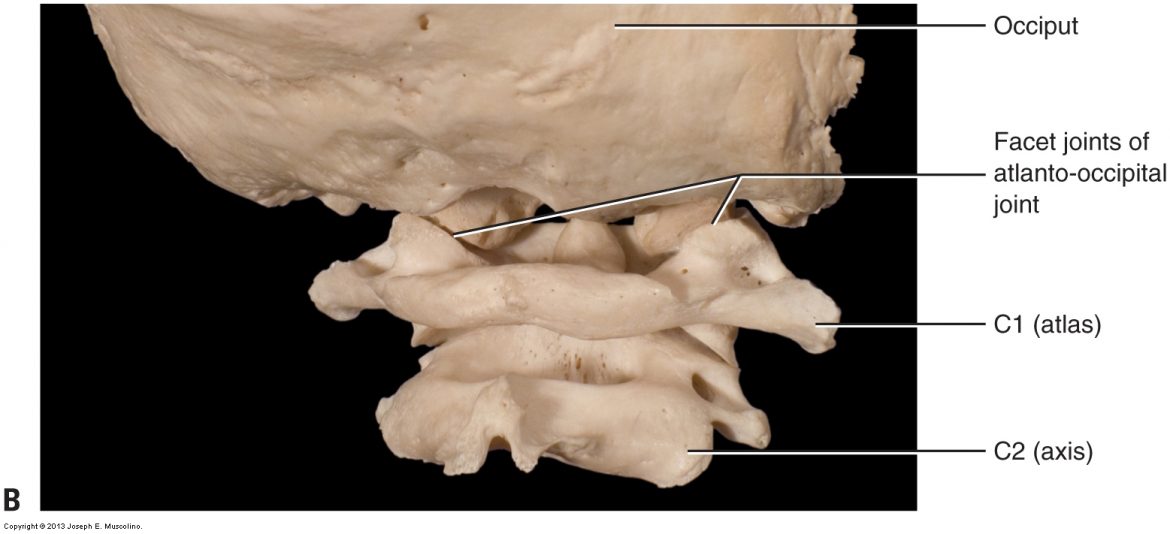
What would have been the body of the atlas is instead fused to the axis, forming the odontoid process (also known as the dens). Therefore, the atlantoaxial (C1-C2) joint, located between the atlas and axis, has no disc joint, nor does the atlanto-occipital (C0-C1) joint, located between the atlas and occiput. The atlantoaxial and atlanto-occipital joints are atypical cervical spinal joints: instead of having two facet joints and one disc joint, the atlantoaxial joint has two facet joints and an atlanto-odontoid joint, and the atlanto-occipital joint has only two facet joints (Fig. 6).
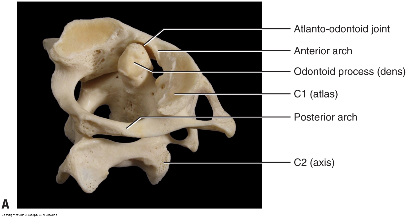

Figure 6. Atlantoaxial and atlanto-occipital joints, posterolateral oblique views. (A) The atlantoaxial (C1-C2) joint located between the atlas and axis. (B) The atlanto-occipital joint located between the atlas and the occiput of the skull. (Courtesy of Joseph E. Muscolino. Photography by David Eliot. Originally published in Kinesiology: The Skeletal System and Muscle Function, 3ed. 2017. Elsevier.)
Cervical Spinal Joints – Facet Joints
Each facet joint is a synovial joint that is located between the inferior vertebra’s superior articular process and the superior vertebra’s inferior articular process (see Fig. 1-5B). It is called a facet joint because the joint surface of each of the articular processes has a facet (a smooth flat surface) on it. The scientific name for facet joints is zygapophyseal joints, hence they are also often known as Z joints. Facet joints function to guide motion at that segmental joint level of the spine. The term segmental joint level refers to a specific joint level of the spine that includes the disc and facet joints at that level (e.g., the joint between C5 and C6, known as the C5-C6 joint, is a segmental level; the C6-C7 joint level is another segmental level).
The disc joint determines how much vertebral motion is possible at a particular segmental level, and the facet joints determine the type of motion (i.e., the direction of motion) that can occur there. At the upper cervical spine, the plane of the facets is perfectly horizontal in the transverse plane. As we descend the cervical spine, the orientation of the facets gradually transitions to be more vertical in the frontal plane. As a general rule, the plane of the cervical facets is usually considered to be an oblique plane with an approximately 45-degree angle between the transverse and frontal planes (Fig. 7A). This angle is often compared to that of the slope of a roof. The type of motion best afforded at each vertebral segmental joint level is important to know and understand when performing joint mobilization (whether it is Grade IV “arthrofascial stretching” or Grade V “chiropractic mobilization / manipulation / adjustment).
Note: Figure 7B is a review of the three cardinal (major) planes of the body. The three cardinal planes are the sagittal, frontal (also known as coronal), and transverse. Any plane that is not perfectly sagittal, frontal, or transverse is an oblique plane.
Because of the orientation of the facets at that level, the upper cervical spine moves extremely well in right rotation and left rotation (transverse plane motions). In fact, nearly half of all the rotation of the entire cervical spine occurs at the atlantoaxial joint. As the planes of the facets of the middle to lower cervical spine transition toward the frontal plane, the lower cervical spine allows less rotation but more lateral flexion. It is especially important to be aware of the type of motion that each region of the cervical spine allows when performing joint mobilization of the neck (discussed in a later blog post). As a rule, the upper neck moves well into rotation, and rotational motion decreases and lateral flexion increases descending the cervical spine.
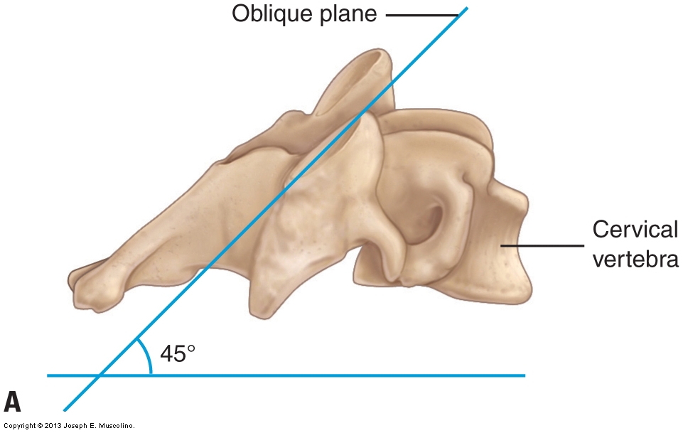
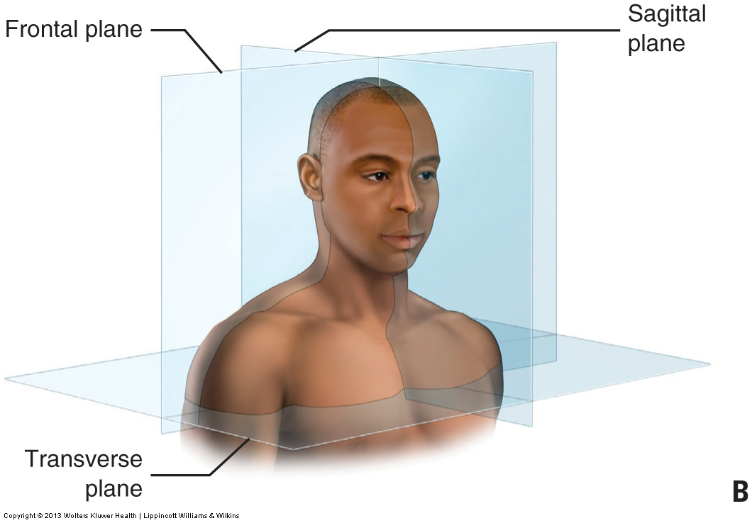
Figure 7. Plane of the facet joints of the cervical spine. (A) The plane of the cervical facets is situated at an approximately 45-degree angle between the transverse and frontal planes. Right lateral view. (Courtesy of Joseph E. Muscolino – Originally published in Advanced Treatment Techniques for the Manual Therapist: Neck. 2013.) (B) Review of all three cardinal (major) planes of the body.
Note: This blog post article is the second in a series of six posts on the
Anatomy / Structure of the Cervical Spine for Manual Therapists.
The Six Blog Posts in this Series are:
- Introduction to the Cervical Spine
- Cervical Spinal Joints
- Motions of the Cervical Spine
- Musculature of the Cervical Spine
- Ligaments of the Cervical Spine
- Precautions When Working the Neck
