
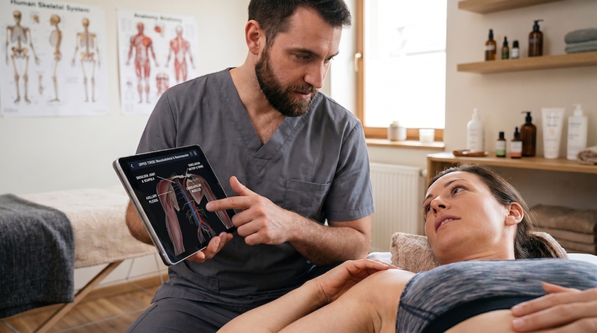
Here’s a scene every hands-on therapist knows. You’ve spent twenty minutes with a client’s shoulder. Your fingers already know the story: where the tissue is stuck, what’s guarding, which movement makes it complain. Then they look up and ask, “So what’s actually going on in there?” And it hits you that the picture so obvious to your hands is completely invisible to them. You point. You say “impingement.” Maybe you wave at a chart taped to the wall. They nod — but it’s a polite nod, not a convinced one. That small disconnect matters more than we usually admit. It quietly decides how much they trust the plan, and whether they actually do their homework between sessions.
The problem isn’t your explanation. It’s their picture.
It’s easy to forget how long it took us to build a working map of the body. Years, really. Clients walk in with none of that. So when you talk about a rotator cuff problem or a cranky lumbar disc, they’re assembling a mental image from scratch, and more often than not it’s stitched together from anxiety and a few half-remembered internet threads.
The usual props only get you so far:
- A wall chart shows “a shoulder.” It doesn’t show their shoulders — layered, tensioned, moving.
- Flat drawings flatten everything. Depth, and the way structures sit against one another, simply disappears.
- And they almost never show the one thing the client actually cares about: why that particular movement hurts.
The result is fairly predictable. The client underestimates what’s happening, or quietly panics, or slowly drifts away from the plan you put together for them.
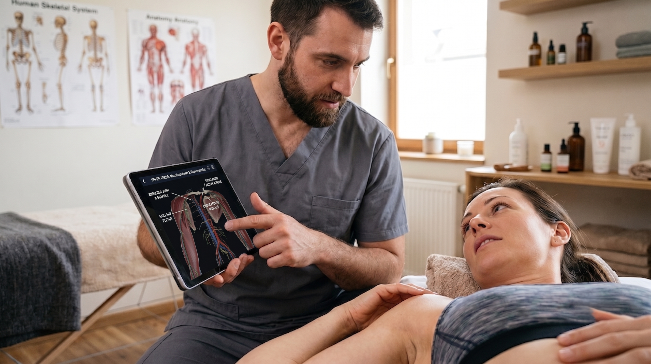
A physical therapist showing an anatomy diagram on a tablet to a female patient
What shifts when the model is in 3D
Hand someone a model they can spin, zoom into, and pull apart layer by layer, and something changes in the room. You stop describing and start pointing at the real thing. Relationships that are nearly impossible to put into words land in a second or two: the way the supraspinatus tucks up under the acromion, how the tissues around your problem spot are all connected to it.
Pathology visualization takes it a step further. You’re not only showing healthy anatomy; you’re setting “normal” right next to the changed version — the worn tissue, the bulge, the inflamed patch. Seeing the two side by side is what turns a scary-sounding label into something a person can actually hold in their head.
From lecture halls to your treatment table
Teaching, and talking shop with colleagues
3D has already changed how the next wave learns anatomy. Students rotate muscles and joints instead of memorizing flat plates, and they come out with a much better feel for palpation and kinesiology. Between practitioners it works as a kind of shared shorthand, too — useful when you’re referring someone out, thinking through a tricky case, or walking a colleague through your reasoning.
But mostly, it’s about the person on your table
The real payoff is smaller and closer than any of that. It’s the client sitting in front of you today. Thirty seconds with a 3D model turns your abstract explanation into something they can point at and still remember on the drive home. This is where a good app earns its keep. Take the VOKA 3D Anatomy & Pathology app: it drops a spinnable atlas of human anatomy, plus a growing set of pathology cases, onto your tablet.
It’s an interactive 3D human anatomy platform built for professionals, the kind of thing you can pull up mid-session to show someone the exact structure you just worked on and the condition you’re treating.
Done briefly and done well, that one moment does a lot of quiet work:
- Trust goes up, because they can see you know precisely what you’re dealing with.
- Compliance improves. Almost nobody skips the home exercise once they’ve actually seen why it matters.
- Expectations get realistic. A recovery timeline makes far more sense after you’ve seen the tissue involved.
- And the fear tends to drop, because a clear picture beats the frightening one they’d been imagining.

A few honest caveats
A 3D model is a teaching aid. It is not a diagnostic tool, and it doesn’t replace clinical judgment or a referral when one is warranted. So a few guardrails are worth keeping in mind:
- Stay in your lane. Use the models to explain and reassure, not to hand out diagnoses that aren’t yours to make.
- Go easy on the drama. A vivid pathology image can frighten someone, or imply more certainty than you actually have. Frame it gently.
- Keep it short. You’re after a lightbulb moment, not a lecture that glazes their eyes over.
And this isn’t going anywhere. As AR and VR keep improving and screen-sharing becomes second nature, these explanations will only get more vivid. The principle underneath stays exactly the same: help the client see what you already understand.
Back to that shoulder
So picture the same client, same question, a few weeks on. This time you turn the screen, spin the model, and show them the structure and the change you’re working on. You watch it land. The nod is different now — that one is understanding, not manners. Your hands were always the skilled part. The 3D just finally lets the client see what you’ve been feeling all along.