
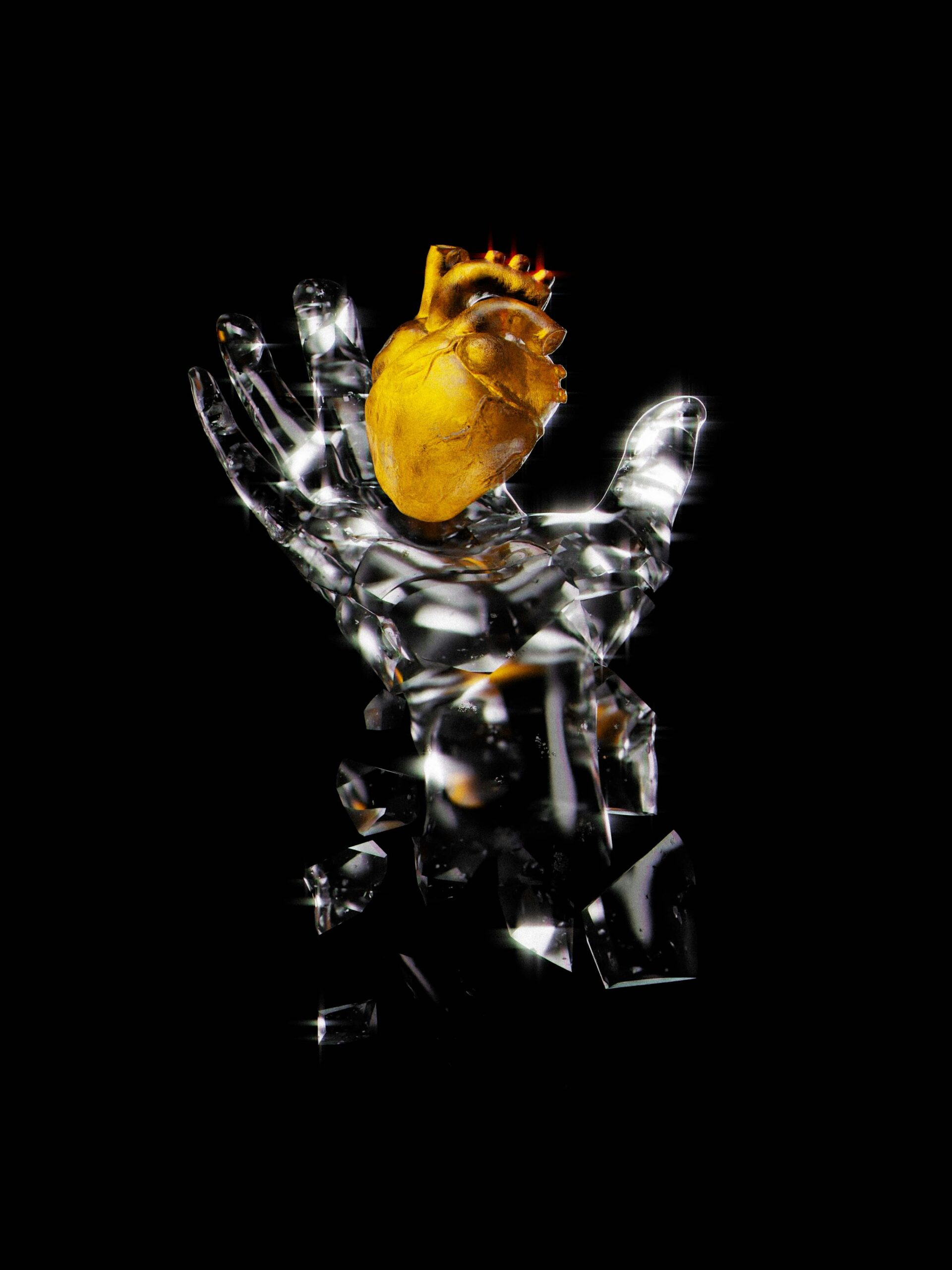
While learning anatomy in college, I quickly realized how formidable it was for students like me to visualize muscles and bones from textbook descriptions. Many of us struggled to understand complex structures from mere words on a page. Even with traditional diagrams, students often couldn’t see what they needed from different angles or for specific scenarios. I knew there had to be a better way!
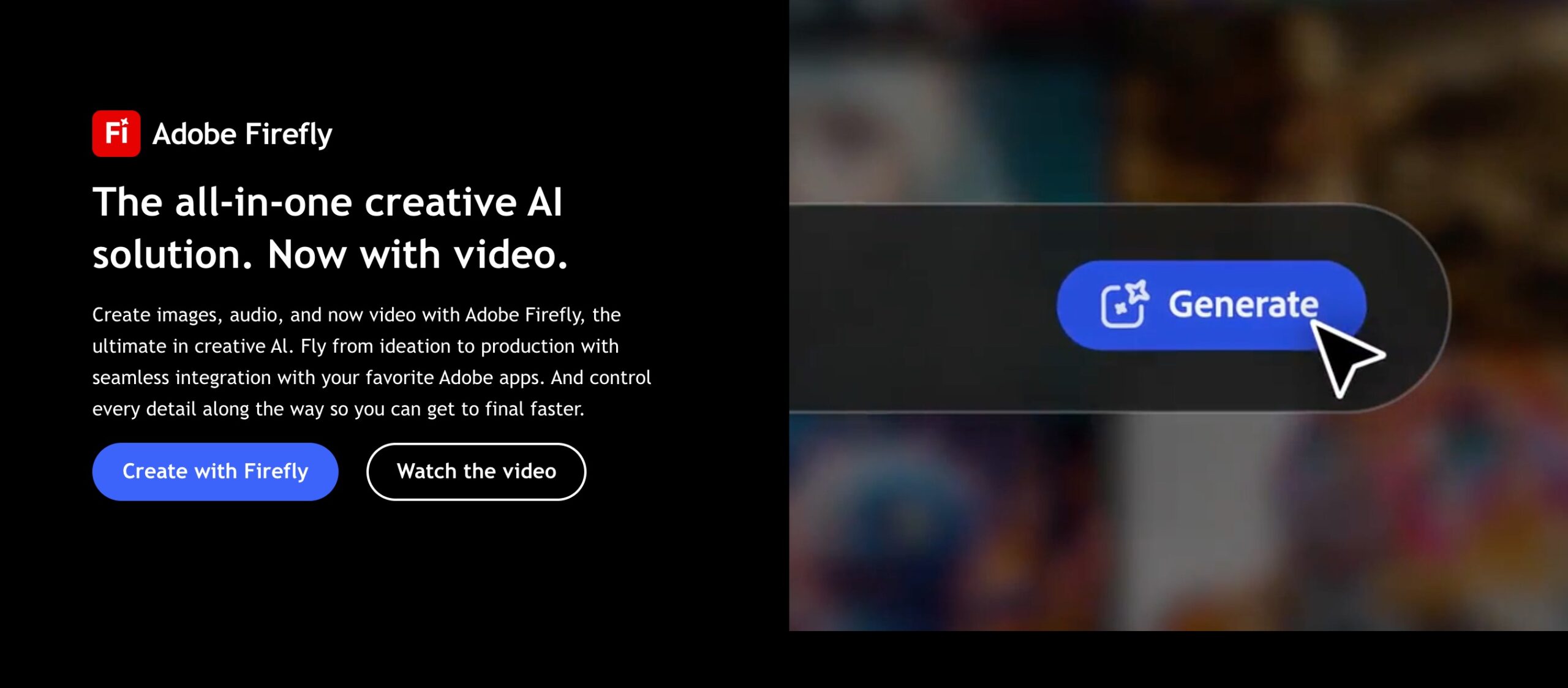
That’s when I discovered how AI technology, like Adobe Firely, could transform anatomy education. Today, teachers and students can create custom muscle illustrations just by typing descriptions. It’s like having a personal medical illustrator available 24/7.
In this article, I’ll show you how this technology works and how you can use it to make learning anatomy easier, more engaging, and more effective!
The Current State of Anatomy Education
If you’ve ever tried to learn or teach anatomy, you know the challenges.
Traditional methods have some real drawbacks:
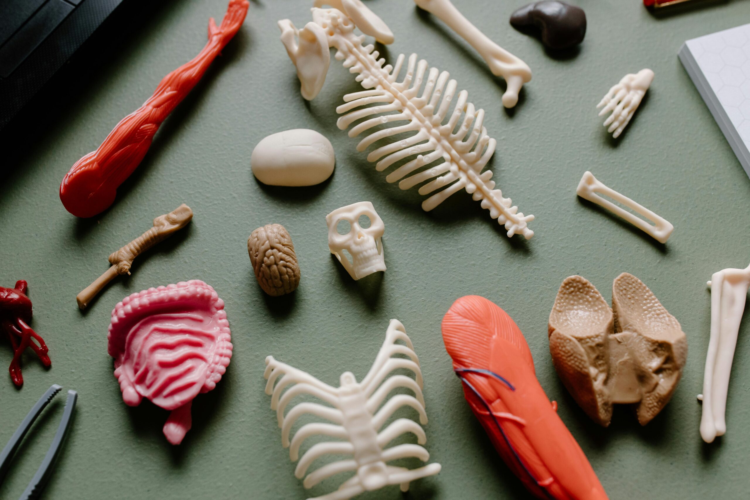
- Textbooks show limited views and can’t be customized
- Professional illustrations are expensive to commission
- Physical models are costly and can’t be modified
- Cadaver labs, while valuable, are limited in availability
Many schools struggle with budget constraints that limit access to high-quality visual resources. Students often feel overwhelmed trying to memorize complex structures without good visual aids. Many of my fellow students easily got frustrated because they couldn’t “see” what they were trying to learn.
Medical and anatomy students typically spend hundreds of hours memorizing structures they can’t visualize well. This is especially true for muscles with complex attachments, actions, and relationships with surrounding structures. When students can’t see these relationships clearly, they often resort to rote memorization, which doesn’t lead to deep understanding.
In my experience studying anatomy, I learn faster and retain information better with strong visual resources. But until recently, creating custom visuals was just too time-consuming and expensive for most educational settings.
How AI-Generated Muscle Illustrations Work
The ability to easily create AI picture generator from text outputs has revolutionized how we can produce detailed, anatomically correct muscle illustrations. The technology behind this is fascinating but not as complicated as you might think.
AI systems like Adobe Firely generate images from text descriptions and operate using neural networks. These networks have been trained on millions of pictures along with their descriptions. For anatomy illustrations, the AI has learned from medical textbooks, anatomical atlases, and other scientific resources.
Here’s a simple breakdown of how it works:
- You type in a description of the muscle or anatomical structure.
- The AI processes your words and understands what you’re asking for.
- It generates an image based on its training.
- You get a custom illustration in seconds.
The key difference between general AI art generators and ones specialized in anatomy is the training data. Anatomy-focused systems have been fine-tuned with medical knowledge, ensuring pristine accuracy in structures, relationships, and terminology.
I recently asked an AI to “show the deltoid muscle from a lateral view with origins and insertions highlighted.” Within seconds, I had an accurate, clear illustration that would have taken a professional artist hours to create.
Benefits for Educators and Students
The advantages of using AI-generated muscle illustrations are game-changing for both teachers and learners:
| For Teachers | For Students |
|
|
When I introduced AI-generated illustrations to younger people studying anatomy, they noticed immediate improvements in their engagement. Students who previously struggled with spatial concepts started asking more insightful questions. They began to understand relationships between structures rather than just memorizing names.
The ability to generate illustrations on demand also means we can create visuals for unusual or difficult-to-find perspectives. If a student is confused about how a particular muscle works during a specific movement, we can generate that exact scenario rather than trying to adapt existing diagrams.
Implementation Guide
If you want to start using AI-generated muscle illustrations in your teaching or learning, here’s how to get the best results:
Creating Effective Prompts
The quality of your illustration depends on how well you describe what you want.
Here are some tips:
- Be specific about the muscle or group (e.g., “brachialis muscle” rather than just “arm muscle”)
- Mention the view you want (anterior, posterior, lateral, medial)
- Specify what you want to be highlighted (origins, insertions, innervation)
- Include context if needed (e.g., “showing relationship to biceps tendon”)
Example Prompts That Work Well:
“Gastrocnemius muscle from posterior view with soleus visible underneath.”
“Rotator cuff muscles with supraspinatus highlighted in red showing impingement.”
“Cross-section of the forearm at mid-level showing muscle compartments and nerves.”
Platforms to Try
- Medical-specific AI image generators (most accurate for anatomy)
- Adobe Firefly for clean, professional illustrations
- General AI art tools with anatomical knowledge
- Educational platforms with built-in AI illustration features
I recommend starting with small, specific requests and gradually working up to more complex illustrations. Always verify the anatomical accuracy by cross-referencing with trusted sources, as AI can occasionally make mistakes.
Examples of Educational Applications of AI-Generated Muscle Illustrations
I’ve seen several impressive applications of AI-generated muscle illustrations in various educational settings:
Physical Therapy Program: A PT instructor used AI illustrations to create custom handouts showing specific muscles involved in common injuries.
Sports Medicine Course: A professor created a series of illustrations showing how the rotator cuff muscles work during a baseball pitch. These custom visuals helped athletes understand injury prevention better than generic diagrams.
Undergraduate Anatomy Lab: Students can create their own AI-generated illustrations as part of their learning process. This activity reinforced their knowledge while teaching them to be precise with anatomical terminology.
Surgical Training Program: A surgical residency program can use AI-generated illustrations to create specialized visuals for pre-operative planning discussions. Residents can now describe specific anatomical variations or surgical approaches they need to visualize and generate custom illustrations showing precisely those scenarios.
Challenges and Limitations
While AI-generated muscle illustrations offer tremendous benefits, they’re not perfect. Here are some challenges I’ve encountered:
- Accuracy Issues: AI occasionally makes anatomical errors, especially with rare or complex structures
- Detail Limitations: Very fine anatomical details may be simplified or missed
- Technical Barriers: Some educators and students may face challenges accessing or using the technology
- Reference Confusion: Without proper guidance, students might trust AI illustrations without verifying the accuracy
Keep in mind that AI-generated illustrations should complement, not replace, traditional anatomical references. They’re tools for learning, not definitive medical references.
Final Thoughts
AI-generated muscle illustrations represent a significant leap forward in anatomy education. They make high-quality, custom visual resources accessible to teachers and students regardless of artistic skill or budget constraints.
In my own learning, I’ve seen how these tools can transform understanding and engagement. Students grasp complex concepts faster and retain information better when they can visualize exactly what they’re learning about.
I encourage you to try these tools in your own teaching or learning process. Start small, be specific with your prompts, and always verify accuracy. The ability to create custom anatomical illustrations on demand is a powerful addition to any anatomy education toolkit.
By embracing these new technologies, we can make anatomy education more accessible, engaging, and effective. That’s something that benefits everyone—teachers, students, and ultimately, the patients who will be cared for by better-educated health professionals.
Written by lawanda@chulasmart.com
