Here’s something every manual therapist eventually learns the hard way: tendons and ligaments just don’t heal like the rest of the body. You can do everything right and a client will still be sitting on your table three months later, still symptomatic, wondering why it’s taking so long. There’s a reason for that, and it’s not a mystery — it’s cellular biology. Once you actually understand what’s happening at that level, a lot of things click into place. How you time your interventions. How you set expectations up front instead of managing disappointment later. How you explain, without sounding like you’re making excuses, why a recovery is taking longer than the client assumed it would.
So that’s what this piece is about — what’s really going on inside a tendon or ligament while it heals, why the process crawls compared to something like a bruise or a cut, and what all of that should mean for how hands-on work gets sequenced through recovery.
Why Tendons and Ligaments Don’t Follow the Usual Rules
Most soft tissue heals in a pretty efficient way. Blood vessels move in, they bring oxygen and repair cells with them, the inflammatory response cleans up the mess, and new tissue gets built in an organized fashion. Muscle is the textbook example — heavily vascularized, so it tends to recover fast.
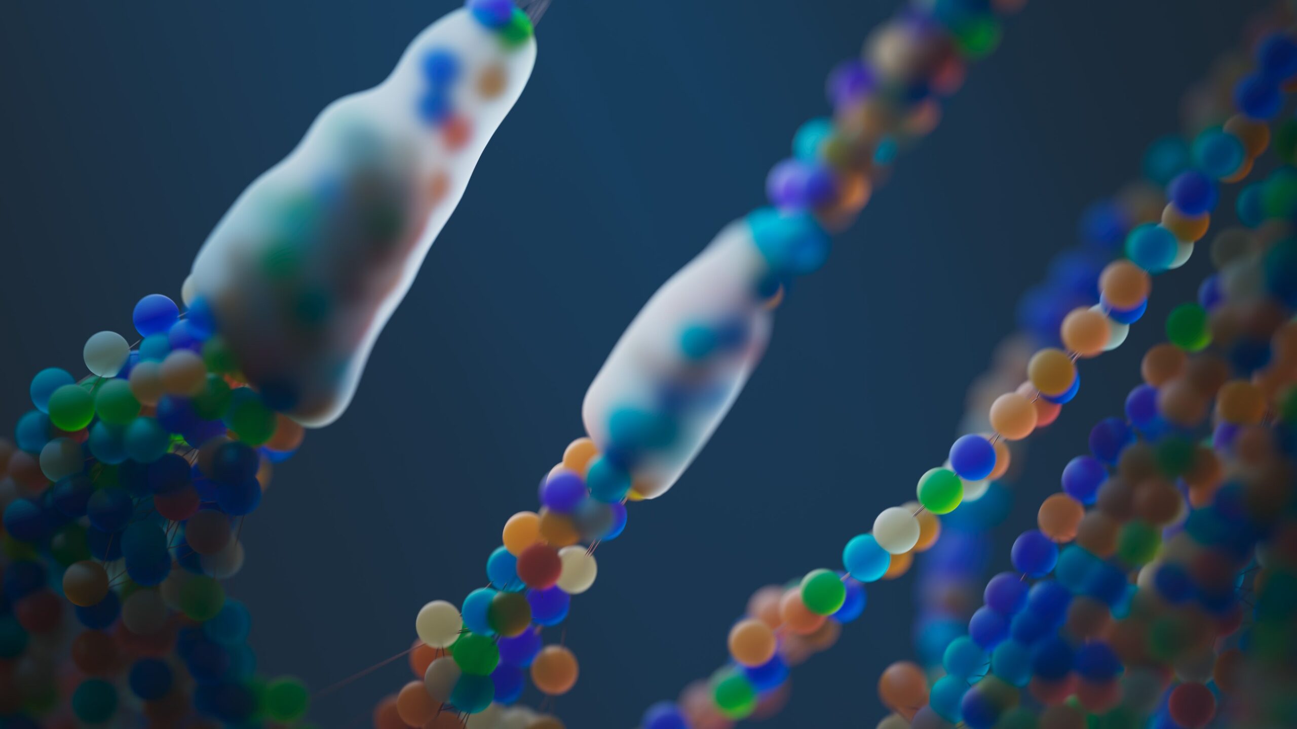
Tendons and ligaments don’t get that luxury. They’re hypovascular — a fraction of the blood supply muscle gets, sometimes far less. And there’s an almost unfair irony baked into their design: the dense collagen that makes them strong enough to hold joints together is the same thing that keeps blood from getting in very deep. Fewer vessels. Fewer repair cells showing up. Less oxygen and fewer nutrients for whatever cells do make it there.
Poor vascularity, on its own, accounts for a lot of what experienced clinicians already sense without needing the biology spelled out. Why a grade II ankle sprain takes so much longer to fully remodel than a similar contusion. Why a partial-thickness rotator cuff tear can still be flaring up months after the “acute phase” is supposedly over on paper.
Three Overlapping Phases, Each With Its Own Rules for Hands-On Work
Repair usually gets broken into three phases in the literature, though in practice they blend into each other more than the diagrams suggest. What matters for us is that each phase calls for a different kind of touch.
Inflammatory phase — roughly days 1 through 5. Mostly vascular and cellular chaos at this point. Platelets clump. Inflammatory cells move in. The body starts hauling out damaged tissue. Hypovascular structures drag this phase out longer than muscle would, purely because the cells doing the work take longer to arrive. Push too hard with manual technique here and you risk stretching the inflammation out further instead of resolving it — which is exactly why most protocols call for protection and gentle, pain-free movement instead of anything deep.
Proliferative phase — roughly days 5 through 21. Fibroblasts run the show now. These are the cells that actually lay down new collagen, and their ability to get into the injury site and start working hinges on angiogenesis — new blood vessel growth. No oxygen, no fibroblast activity, essentially. Tendon and ligament tissue is noticeably slower to grow these new vessels than muscle is, and that alone stretches the proliferative phase out across weeks rather than days. What gets laid down early is disorganized, mostly Type III collagen — weaker, less mature than the Type I collagen it’ll eventually be replaced by.
Remodeling phase — weeks to months, occasionally well over a year for the bigger ligament structures. This is where the messy collagen from the proliferative phase gets reorganized along actual lines of mechanical stress. It’s also the phase where your hands-on work and progressive loading have the biggest say in the outcome. Controlled mechanical stress — through the right manual technique combined with graded exercise — is honestly one of the main things driving how those collagen fibers end up aligned. Skip the mechanical input and the tissue tends to remodel into something weaker and less organized. That’s really the whole case for pairing manual work with active rehab instead of leaning on just one or the other.
Why This Matches What You Actually See in the Clinic
Two patterns clinicians run into constantly stop being mysterious once the vascularity piece is understood.
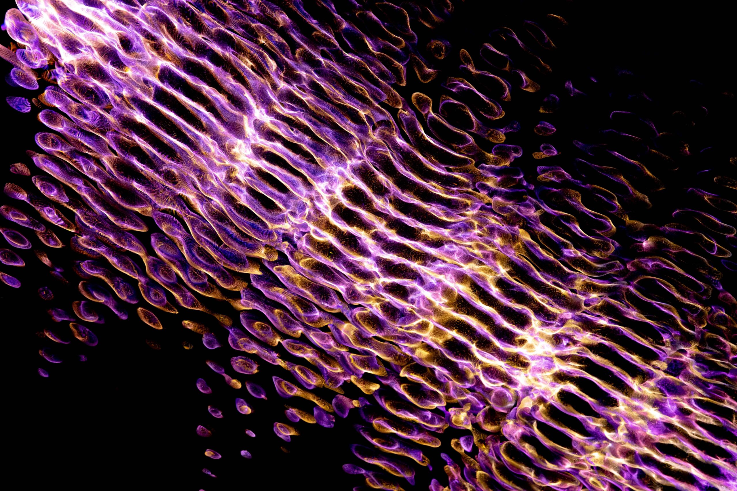
One is why tendinopathy shows up so often as a chronic, low-grade nuisance instead of a clean acute injury. Slow, uneven healing means a tendon can pile up microtrauma faster than it can actually repair it — and what ends up on imaging is that degenerative, disorganized collagen pattern typical of chronic tendinopathy, not anything resembling a tidy inflammatory response.
The other is why loading progressions for ligament injuries move so much slower and more cautiously than they would for a muscle strain of similar severity. It’s not caution for its own sake — the biology underneath is genuinely running on a different clock, and no amount of client motivation speeds that up. Anyone wanting a deeper technical dive into how vessel formation drives this specific timeline can check out this overview of angiogenesis and tendon repair mechanisms.
An Active, Evolving Corner of Research
Hypovascularity and sluggish fibroblast recruitment are basically the two bottlenecks holding tendon and ligament healing back — which is exactly why so much current research attention is pointed at compounds that interact with those two processes specifically. It’s not a coincidence that peptide research keeps circling back to angiogenesis and fibroblast activity in tendon models. Those are the mechanisms sitting closest to the actual problem.
Take BPC-157 as one example — it’s been studied in animal models for how it interacts with VEGF signaling (vascular endothelial growth factor, one of the main drivers behind angiogenesis), and researchers have documented effects on tendon-to-bone healing and collagen organization in preclinical settings. A comparison of how different research peptides approach tissue repair is worth a look too, since it shows just how many different angles this research takes — vascular signaling, cell migration, collagen synthesis, each compound tackling the problem from a different direction. Worth being upfront that this is still lab-stage, evolving research, not something that’s crossed over into clinical practice. But it says something that so much of it circles right back to the same two bottlenecks — vascularity and fibroblast recruitment — that show up clinically every day. Readers interested in the current state of this research can find a broader overview of the compounds being studied in tissue repair research.
What to Actually Carry Into Practice
Understanding the cellular timeline gives you something more solid to build a treatment plan around than just following a generic protocol because that’s what the protocol says. Some things worth keeping in mind:
- Hypovascular tissue is going to run a longer inflammatory phase than muscle would — plan your intervention intensity around that, not around the calendar.
- Early proliferative-phase collagen is weak and disorganized. Symptoms settling down doesn’t mean the tissue is ready for aggressive loading — those two things aren’t the same signal.
- Treat the remodeling phase as deliberate work, not just “time passing.” Mechanical stress is genuinely doing something to how that collagen organizes itself, which is the real argument for pairing manual technique with a structured loading plan rather than picking one over the other.
- When a client’s expectations don’t line up with where the tissue actually is, walking them through why hypovascular tissue behaves differently than muscle tends to land better than just asking them to be patient.
Tissue repair isn’t one event with a start and finish line. Tendons and ligaments run on a different clock than the rest of the musculoskeletal system, and keeping that in view is what turns a generic treatment timeline into one that’s actually built around how the tissue in front of you is going to heal.
Author: Sarah Whitfield Sarah writes on musculoskeletal biology and tissue repair research for Patriot Peptides, focusing on the cellular side of soft tissue healing and where peptide research currently stands. She’s especially interested in taking dense physiology and turning it into something clinicians and researchers can actually use.
Email: patriotpeptides@gmail.com