Achilles Tendon

Posterior view of the right leg. Permission Joseph E. Muscolino. The Muscular System Manual – The Skeletal Muscles of the Human Body, 4ed (Elsevier, 2017).
The Achilles tendon is the largest tendon in the human body, it consists of sub-tendons from 3 muscles/muscles bellies of the triceps surae group (soleus, lateral gastrocnemius, medial gastrocnemius). These sub-tendons together form the tendinous part of the Achilles tendon and twist as they descend into the distal attachment (insertion) at the calcaneal bone. The Achilles tendon handles functional loads applied to the tendon. It also balances resisting tension and allowing compliance, as the function of tendons is more complex than just transmitting force from muscle to bone. They store and release energy, protect muscle from stretch damage, allow favorable muscle output and enhance muscle performance.
Twisted Structure Study
A study from Poland published in Scandinavian Journal of Medicine & Science in Sports investigated the twisted structure of the Achilles tendon and its individual sub-tendons. Specimens of the Achilles tendon, with preserved calcaneal bone and a fragment of the triceps surae musculature, were obtained from 53 fresh-frozen, male cadavers (n=106 lower limbs).
The twisted structure of the Achilles tendon has been described as a clockwise rotation in the left limb and counter clockwise rotation in the right limb (as seen from proximal to distal end).
Results of Study
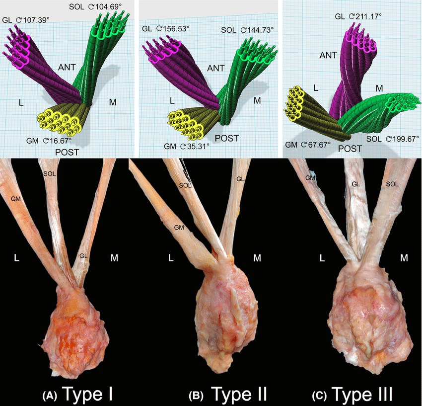
Types of the left Achilles tendon torsion (top: three-dimensional model, below: cadaveric pictures: dorsal view). ANT, anterior; POST, posterior; L, lateral side; M, medial side; GL, the sub-tendon originating from the lateral head of gastrocnemius muscle; GM, the sub-tendon originating from the medial head of gastrocnemius muscle; SOL, the sub-tendon originating from the soleus muscle (From Pekala et al. 2017).
This study posits that there are three types of the Achilles tendon with differently twisting sub-tendons:
- Type I—Least twisted, in which the part of the Achilles tendon originating from the gastrocnemius muscle is attached to the posterior two-thirds of the calcaneal tuberosity, and the soleus tendon is attached to the posterior one-third of the calcaneal tuberosity. This was the most prevalent form, found in 52% of the subjects.
- Type II—Moderately twisted, in which the part of the Achilles tendon originating from the gastrocnemius muscle is attached to the posterior half of the calcaneal tuberosity, and the tendon from the soleus is attached to the other half of the calcaneal tuberosity. This was the second most prevalent form, found in 40% of the subjects.
- Type III—Extremely twisted, in which all three sub-tendons of the Achilles tendon (medial head of the gastrocnemius, lateral head of the gastrocnemius, and soleus muscle) have a twisted structure. This was the least common form, found in 8% of the subjects.
The authors also found that the torsion angle of the Achilles tendon’s superficial (posterior) fibers (medial gastrocnemius) is significantly smaller than that of the fibers lying deeper (more anteriorly) in the tendon (lateral gastrocnemius and soleus). The greatest degree of Achilles tendon torsion is observed in the deepest (most anterior) part of the tendon (lateral gastrocnemius and soleus).
Mechanism
The authors concluded that types of Achilles tendon torsion may potentially alter the biomechanical properties of the tendon, thus possibly influencing the pathophysiologic mechanisms leading to the development of various tendinopathies.
Another Study
Another study from Belgium also showed that the Achilles tendon displayed a twisted structure, with the greatest twisting found in the deep (anterior) layer of the tendon (lateral gastrocnemius and soleus). Further, a non-uniform regional strain behavior was found in the Achilles tendon during passive elongation, with the highest strain in the superficial layer.
This blog post article was created in collaboration with www.terrarosa.com.au.
(Click here for the blog post article: How Do We Treat Achilles Tendon Disorders with Manual Therapy.)
