Fibrosus versus Densification
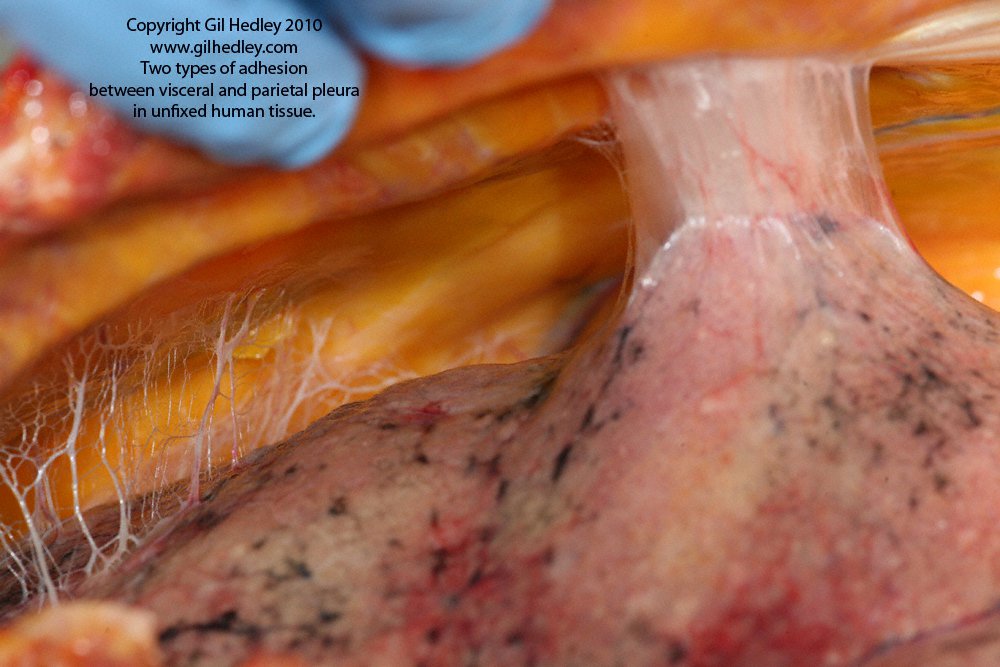
Densification on the left. Fibrosus on the right.
Copyright: Gil Hedley (https://www.gilhedley.com/).
Deep fascia has been considered one of the origins of pain and dysfunction. The terms “fibrosis” and “densification” are often used to indicate changes in the deep fascia. They can modify the mechanical properties of the deep fascia and damage the function of underlying muscles or visceral organs. However, the two terms refers to two different alterations of fascia.
Authors Dr. Piero G. Pavan, Dr. Antonio Stecco, Dr. Robert Stern, and Dr. Carla Stecco wrote a review in Current Pain & Headache Report (2014).
The authors said that understanding the connective tissue matrix within fascia and distinguishing between these two different alterations in fascia will enable the prescription of more specific manual and movement treatment modalities to relieve chronic pain syndromes.
Deep Fascia
Deep fascia, in this case, refers to any dense fibrous sheath that interpenetrates and surrounds the muscles, bones, nerves, and blood vessels of the body, binding all these structures together into a firm, compact mass. Over bones, it is called periosteum; around tendons, it forms the paratendon; around vessels and nerves, it forms the neurovascular sheath; around muscles it forms intermuscular septa and other fascial sheaths; and around joints, it strengthens the capsules and ligaments.
Deep fascia (such as fascia latae, brachial fascia, crural fascia) is not a single structure, but a multilayer structure composed of two to three layers of dense connective tissue (collagen type 1, 3, elastin fibers), and several layers of loose connective tissue (adipose tissue, GAG [glycosaminoglycan], and HA [hyaluronic acid]).
Dense Deep Fascia
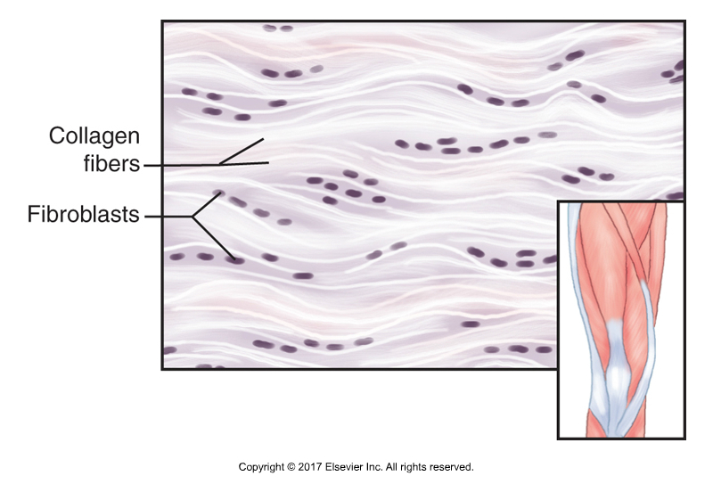
Dense deep fascia. Permission: Joseph E. Muscolino. Kinesiology – The Skeletal System and Muscle Function, 3rd Edition, 2017. Elsevier.
The dense connective tissue has a function to transmit force, proximally to distally and vice versa, and to help the coordination of muscles. We know now that about 30% of the muscle attachments are not in the tendon, but into the deep fascia. All of these fascial attachments provide an excellent illustration of how the thickness and strength of fascia is a precise mirror of the forces generated by muscular action. When these muscles contract, not only do the bones move, they also pull on and transmit forces into the deep fascia.
Loose Deep Fascia
Meanwhile, the loose connective fascial tissue is structured so that it is independent of the deep fascia. Loose fascial tissue allows gliding between layers of deep fascia and between layers of deep fascia and adjacent structures such as musculature.
Deep fascia is also innervated with many mechanoreceptors (proprioceptors such as Ruffini and Pacini receptors, and free ending nerves). Research from Heidelberg led by Siegfried Mense also described that fascia is innervated with nociceptors (pain receptors). Fascia is therefore also important in proprioception and pain generation. It is possible that the viscoelasticity of fascia can modify activation of the nervous system receptors within fascia.
Digital COMT
Did you know that Digital COMT (Digital Clinical Orthopedic Manual Therapy), Dr. Joe Muscolino’s continuing education video streaming subscription service for massage therapists (and all manual therapists) and movement professionals, has six folders with video lessons on Manual Therapy Treatment, including an entire folder on Pathomechanics, as well as a folder on Anatomy, and many, many more on manual and movement therapy techniques? Digital COMT adds seven new video lessons each and every week. And nothing ever goes away! Click here for more information.
Alterations of the Deep Fascia
The authors postulated that the deep fascia could be subjected to at least two different kinds of alterations:
(1) Densification: Alteration of the loose component that affects the sliding system between fascial tissue interfaces.
(2) Fibrosus: Alteration of the fibrous collagen component that affects the capacity of load transmission.
“Fibrosis” is similar to the process of scarring, with the deposition of excessive amounts of dense fibrous collagen connective tissue, reflective of a reparative or reactive process. It can obliterate the architecture and function of the involved fibrous tissue.
On the other hand, “densification” refers to an increase in the density of fascia, affecting its loose fascial component, but maintaining the architecture of the fibrous fascias tissue and therefore its function. Because the loose fascial component of deep fascia is concerned with the ability of deep fascial tissue to glide along adjacent layers of tissue, densification is a modification of the mechanical gliding proprieties of fascia, without altering the general architectural structure of its collagen framework.
Densification results in increased viscosity of the loose connective tissue within fascia and can be caused by a concentration of hyaluronic acid. Hyaluronic acid occurs between deep fascia and muscle, facilitating gliding between these two structures, and also within the loose connective tissue of the fascia, guaranteeing the smooth sliding of adjacent fibrous fascial layers. Densification does not alter collagen fiber bundles but can diminish the gliding between adjacent fibrous layers and therefore change the tensile status of the fascia.
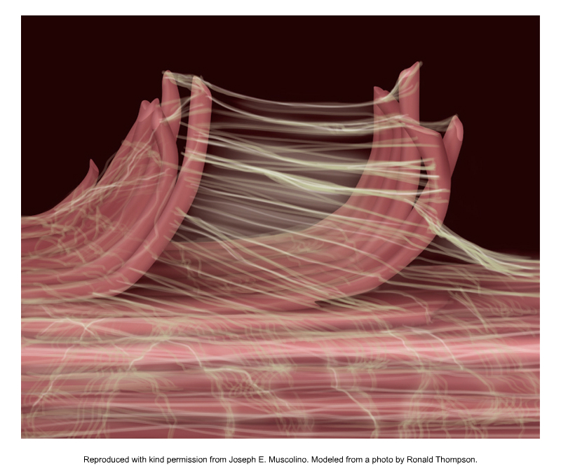
Myofascial tissue. Permission: Joseph E. Muscolino. Kinesiology – The Skeletal System and Muscle Function, 3rd Edition, 2017. Elsevier. Modeled on a photo by Ronald Thompson.
There are several factors that affect densification and fibrosis:
- Diet, exercise, and overuse syndromes that cause an alteration of the loose connective tissue inside the deep fascia, causing fascial densification. This alteration is easily reversible because we can modify the mechanical properties of the extracellular matrix by increasing the temperature, or increasing local strain with a mechanical stimulus, such as massage therapy.
- Trauma (including overuse trauma), surgery, and diabetes can alter the fibrous layers of the deep fasciae, causing fascial This alteration is difficult to modify once formed because only a local inflammatory process can destroy the pathological collagen fibres and permit deposition of new collagen fibres. Only early guided mobilization allows healing of the deep fascia to avoid the formation of fibrosis.
Studies Concerning Altered Deep Fascia
Work by Helene Langevin and colleagues investigated the sliding capability of dense layers. They found significant correlations in people with chronic low back pain between shear strain capability of the loose connective layer and the following variables: perimuscular connective tissue thickness, echogenicity, trunk flexion range of motion, and trunk extension. Another study on animal demonstrated that an induced soft tissue injury in the lumbar region, when combined with movement restriction, lead to fibrosis, and significant thickening of thoracolumbar fascia. These studies demonstrate the importance of altered sliding of the thoracolumbar fascial layers in low back pain.
Antonio Stecco and colleagues showed a relationship between a decrease in the range of motion and increases in the thickness of neck deep fasciae. Using ultrasonography, the authors found a mean thickness of fascia of 1.1 mm of the sternocleidomastoid (SCM) muscle in healthy participants, while patients with chronic, nonspecific neck pain have a mean fascia thickness of 1.8 mm. In the healthy participants, the deep fascia of SCM appears as a white, fibrous layer, while in subjects with neck pain, the sublayers forming the deep fascia are recognized by the increased thickness of the loose connective tissue.
Comment by Joseph E. Muscolino
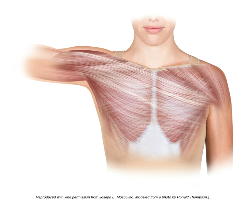
Fascia responding to mechanical forces and increasing its integrity. Permission: Joseph E. Muscolino. Kinesiology – The Skeletal System and Muscle Function, 3rd Edition, 2017. Elsevier.
Deep fascia, like all tissues of the body, can respond to the forces placed upon it as well as the chemical and nutritional quality of the surrounding extracellular matrix. When the forces placed upon the deep fascia are a challenge to the tissue but graded in intensity and not too severe, the fascia can respond by increasing its fascial collagen structure and therefore increasing its integrity. Similar to how muscle tissue can adapt to the increased demands of exercise and become stronger and healthier, so can fascia.
However, when the forces placed upon the fascia are too great for the tissue to respond to in a healthy manner, the deep fascia can be altered in two manners: fibrosis and densification. Fibrosis could be considered to be a pathologic process that affects the structural architecture of fibrous collagen tissue and therefore diminishes the proper functioning of fascia to absorb and transmit tensile (pulling) forces. Densification, on the other hand, can be viewed as an alteration in the loose fascial tissue components of deep fascial. This can impact the ability of the deep fascial tissue layers to glide along adjacent tissue interfaces (such as alone adjacent muscle or bone). Then, because of fascia’s role in proprioception and pain, these alterations may lead to greater pain and dysfunctional patterns in the body.
Between the two types of alterations of deep fascia, densification is the easier to treat and reverse with such manual and movement techniques as massage therapy, stretching, joint mobilization, and exercise. Fibrosus, on the other hand, once it has formed, because it is the effective accumulation of dense fibrous collagen, is much more resistant to manual and movement therapies and tends to become a more chronic problem.
It is a time-worn cliché but true statement: “An ounce of prevention is worth a pound of cure.” Given the challenge to treating fibrosus, our goal as manual therapists and movement professionals should be to guide our patients and clients toward a lifestyle that prevents the dysfunctional alteration to fascial structure such as densification, and especially, fibrosus.
This blog post article was created in collaboration with terrarosa.com.au.
(Click here for the blog post article: Intermuscular Force Transmission Along a Myofascial Chain.)
Digital COMT
Did you know that Digital COMT (Digital Clinical Orthopedic Manual Therapy), Dr. Joe Muscolino’s continuing education video streaming subscription service for massage therapists (and all manual therapists) and movement professionals, has six folders with video lessons on Manual Therapy Treatment, including an entire folder on Pathomechanics, as well as a folder on Anatomy, and many, many more on manual and movement therapy techniques? Digital COMT adds seven new video lessons each and every week. And nothing ever goes away! Click here for more information.
