Overview of New Findings – The Lateral FibuloTaloCalcaneal Ligament

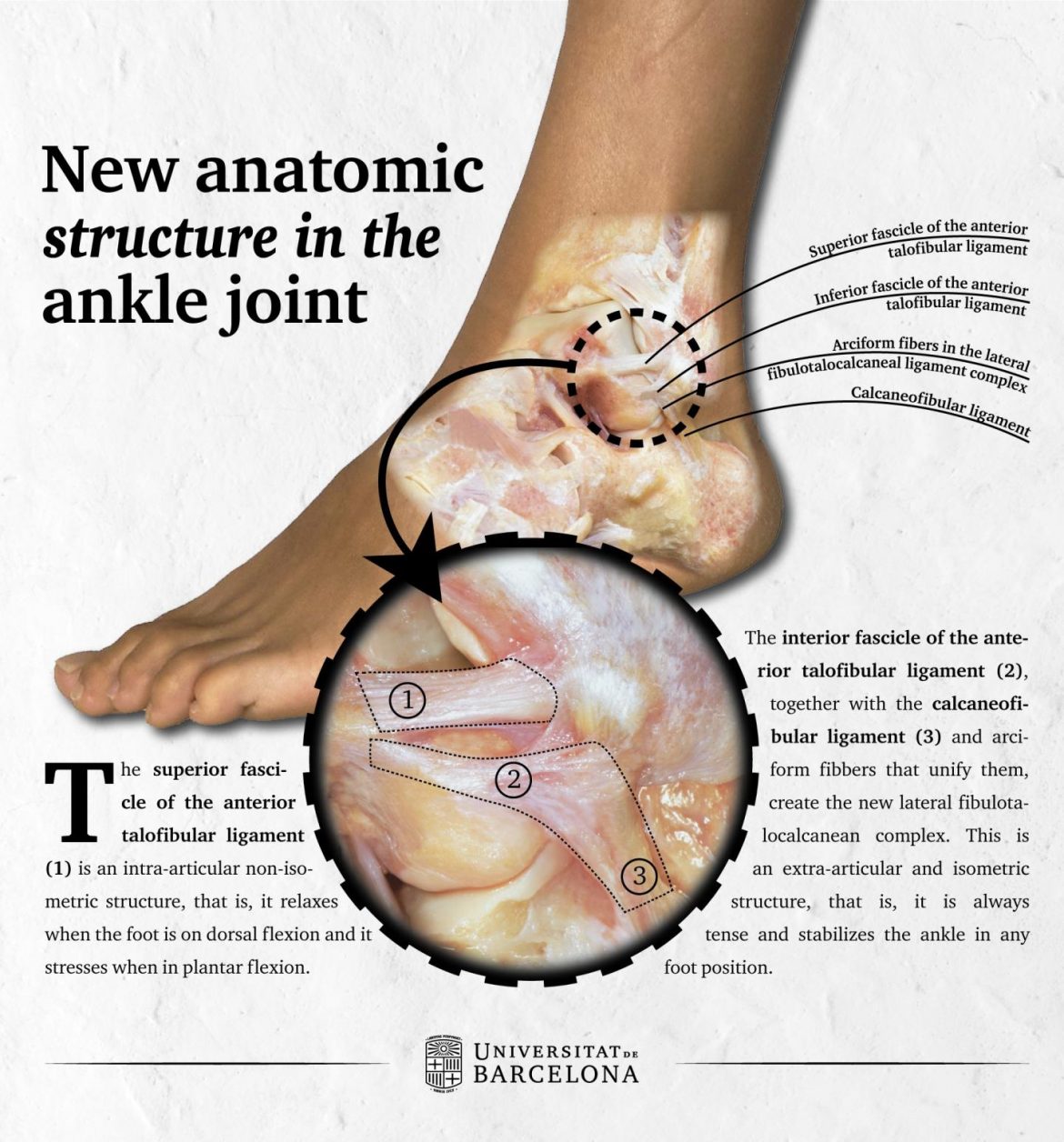
The Lateral Fibulotalocalcaneal Ligament. The superior fascicle of the anterior talofibular ligament (1) is an intra-articular ligament. The inferior fascicle of the anterior talofibular ligament (2), together with the calcaneofibular ligament (3), and arciform fibers that unite them, create the new lateral fibulotalocalcaneal ligament; this ligament is extra-articular.
Recent analysis of the lateral collateral ligament complex – anterior talofibular ligament, calcaneofibular ligament, and posterior talofibular ligament – has yielded some new findings. Following is an overview of these findings:
- There are ligament fibers that connect the anterior talofibular ligament to the calcaneofibular ligament. These connecting fibers are described as arciform because of they are arc-shaped.
- Therefore, the anterior talofibular ligament and the calcaneofibular ligament, along with the connecting fibers, have been named as one structure: the lateral fibulotalocalcaneal ligament (Lateral Fibulo-Talo-Calcaneal Ligament) (LFTCL). In other words, one ligament on the lateral side of the ankle joint that connects the fibula to the talus and calcaneus.
- The old-named anterior talofibular ligament is actually composed of two distinct sets of fibers (fascicles): the superior anterior talofibular ligaments and the inferior anterior talofibular ligament.
- The superior anterior talofibular ligament is actually intra-articular, meaning that it is located within the ankle joint.
- The inferior talofibular ligament is extra-articular (outside of the joint) and is the fascicle of fibers that is connected to the calcaneofibular ligament, and therefore these two ligaments are really one functional unit and newly named as the fibulotalocalcaneal ligament.
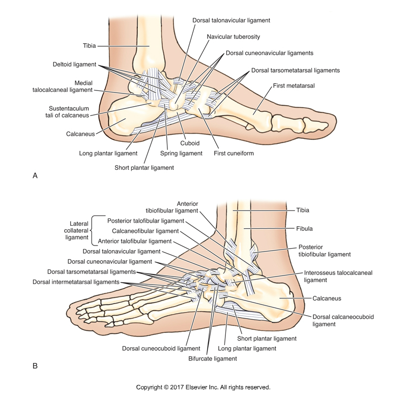
Classic description of the ligaments of the ankle joint. A, Medial view. B, Lateral view. Permission: Joseph E. Muscolino. Kinesiology – The Skeletal System and Muscle Function, 3rd Edition (Elsevier, 2017).
Lateral Collateral Ligament Complex
Got all this?… So now the lateral collateral ligament complex is still composed of three parts, but they are now divided as follows:
- Superior anterior talofibular ligament
- Fibulotalocalcaneal ligament (inferior anterior talofibular ligament and calcaneofibular ligament)
- Posterior talofibular ligament
Did you know that Digital COMT (Digital Clinical Orthopedic Manual Therapy), Dr. Joe Muscolino’s continuing education video streaming subscription service for massage therapists (and all manual therapists and movement professionals), has at present (November of 2018) more than 1,000 video lessons on manual therapy continuing education, including entire folders on stretching, joint mobilization, and massage. And we add seven (7) new videos lessons each and every week! And nothing ever goes away. There are also folders on Anatomy and Physiology, including an entire folder on Cadaver Anatomy… and many, many more on other manual and movement therapy assessment and treatment techniques. Click here for more information.
Study
Although the lateral collateral ligament complex of the ankle joint had been well studied, there were no specific descriptions of whether or how these ligaments are connected to each other as part of the same complex. Therefore, a study was conducted by researchers at the University of Barcelona. This study was published in the Knee Surgery, Sports Traumatology, Arthroscopy journal.
The researchers conducted an anatomical study on 32 fresh-frozen below-the-knee ankle specimens. A plane-per-plane anatomical dissection was performed. The characteristics of the anterior talofibular ligament and calcaneofibular ligament, as well as any connecting fibers between them were recorded. Measures were obtained in plantarflexion and dorsiflexion, and by two different observers.
Results
The observation showed that the anterior talofibular ligament in all the specimens is a two-fascicle ligament (composed of two distinct sets of fibers and really two distinct ligaments). These two distinct parts can be described as the superior anterior talofibular ligament and the inferior anterior talofibular ligament. Further, the superior anterior talofibular ligament fascicle was observed to be intra-articular within the ankle joint, in contrast to the inferior anterior talofibular ligament, which is extra-articular (outside of the ankle joint).
The fibers link the inferior fascicle of the anterior talofibular ligament and the calcaneofibular ligament. This connection had never been described before, and it suggests that both ligaments are truly one functional unit, therefore renamed together as the lateral fibulotalocalcaneal ligament (or the lateral fibulotalocalcaneal ligament complex).
Isometric and Non-Isometric Ligaments
Further, the authors of this study made the following statements:
- The distinct superior anterior talofibular ligament is a non-isometric ligament, meaning that it can be taut or relaxed. It is taut when the ankle joint is in plantarflexon, whereas it is relaxed when the ankle joint is in dorsiflexion.
- The newly named lateral fibulotalocalcaneal ligament is an isometric ligament, meaning that it is always taut, regardless of the position of the ankle joint.
Clinical Implications
The clinical implications of this study are:
- The superior fascicle of the anterior talofibular ligament is anatomically a distinct structure from the inferior anterior talofibular ligament fascicle; therefore they are somewhat functionally different from each other (meaning that one fascicle versus the other fascicle can be taut or relaxed in different positions of the foot).
- The superior fascicle is an intra-articular ligament, that will most probably not be able to heal after a rupture, that is why it is so frequent for patients to have chronic symptoms after an ankle sprain, especially if an isolated rupture of this fascicle is present.
- This has implications in the surgical treatment of ankle sprains.
- Development of clinical tests is needed to assess an injury of the superior anterior talofibular ligament from an injury of the lateral fibulotalocalcaneal ligament.
This blog post article was created in collaboration with www.terrarosa.com.au.
Digital COMT
Did you know that Digital COMT (Digital Clinical Orthopedic Manual Therapy), Dr. Joe Muscolino’s continuing education video streaming subscription service for massage therapists (and all manual therapists and movement professionals), has at present (November of 2018) more than 1,000 video lessons on manual therapy continuing education, including entire folders on stretching, joint mobilization, and massage. And we add seven (7) new videos lessons each and every week! And nothing ever goes away. There are also folders on Anatomy and Physiology, including an entire folder on Cadaver Anatomy… and many, many more on other manual and movement therapy assessment and treatment techniques. Click here for more information.
