Epimuscular Myofascial Force Transmission
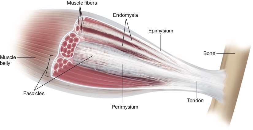
Muscular fascia. Permission Joseph E. Muscolino. www.learnmusles.com. Art work by Giovanni Rimasti.
Epimuscular myofascial force transmission is a concept that concerns the transmission of muscular forces to the skeleton via pathways other than the typically thought of mechanism of muscle force transmission created by the sliding filament mechanism.
Sliding Filament Mechanism for Myofascial Force
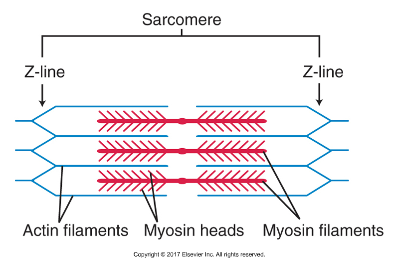
The sliding filament mechanism is the mechanism with the sarcomere (sarcomeres are units of muscle tissue) whereby myosin filament heads form cross-bridges with active binding sites on actin filaments, and then creating pulling forces that attempt to pull the actin filaments toward the center of the sarcomere. If the actins are pulled in toward the center, then the Z-lines (borders) of the sarcomeres are pulled closer together and the sarcomere shortens. When sarcomeres in series (laid end to end) all shorten, the myofibril in which they are located shortens. When myofibrils of the muscle fiber all shorten, the muscle fiber shortens. When enough muscle fibers shorten, the muscle shortens and pulls and moves one or both of its (bony) attachments toward the other. This is essentially how a shortening concentric muscle contraction occurs.
Note:
- This typical model for muscle force transmission via the sliding filament mechanism can be termed intramuscular myofascial force transmission(“intra” means inside).
- Therefore, epimuscular myofascial force transmission that this article is about can also be called extramuscular myofascial force transmission (“extra” means outside).
- Epimuscular myofascial force transmission can also be called intermuscular myofascial force transmission(“inter” means between).
- Although some sources reserve the term extramuscular myofascial force transmissionfor when the transfer of force occurs between a non-muscle tissue (such as an interosseus septum, ligament, or neurovascular bundle) and a muscle.
- And these sources reserve the term intermuscular myofascial force transmissionfor when the force transfers from the movement of one muscle to another muscle (in other words, between muscles given that “inter” means between).
- AND… Another term used to describe the ability of one muscle to affect the force of another adjacent muscle is lateral force transmission.
- Short comment by Joseph Muscolino: “Lots of terms, eh? Don’t feel bad if you need to read this blog post article a few times before getting it all. I had to read the study a number of times before I could write this! See my comment / summation below for the point of all this.”
Permission Joseph E. Muscolino. Kinesiology – The Skeletal System and Muscle Function, 3rd ed. (Elsevier, 2017).
Epimuscular Myofascial Force Transmission
Epimuscular myofascial force transmission is different from this classic view of skeletal muscle contraction force transmission, where force is generatedwithin its muscle fibers (sarcomeres) and then directly transmitted in-series, usually via a tendon, onto the skeleton. While there are many studies on animals and human cadavers that prove the existence of epimuscular myofascial force transmission, study on living human is still lacking.
Digital COMT
Did you know that Digital COMT (Digital Clinical Orthopedic Manual Therapy), Dr. Joe Muscolino’s continuing education video streaming subscription service for massage therapists (and all manual therapists and movement professionals), has at present (December of 2018) more than 1,000 video lessons on manual therapy continuing education, including entire folders on massage therapy, stretching, and joint mobilization. And we add seven (7) new videos lessons each and every week! And nothing ever goes away. There are also folders on Pathomechanics and Anatomy and Physiology, including an entire folder on Cadaver Anatomy… and many, many more on other manual and movement therapy assessment and treatment techniques? Click here for more information.
Study
A studyfrom Japan examined if manipulating joint angle to stretch the muscle can alter the shear modulus of an adjacent restingmuscle. The biceps brachii (manipulated muscle) and the brachialis (resting adjacent muscle) were the focus of this study because they are neighbouring muscles, yet have independent tendons that essentially insert onto different bones (biceps brachii onto the radius and brachialis onto the ulna).
Thirteen healthy young adult men participated in the study. To manipulate the muscle length of biceps brachii only, the radius of the forearm was passively setat supination, neutral, and pronation positions (the brachialis attaching onto the ulna and not the radius did not have its length changed with movement of the radius). The shear modulus (muscle stiffness) of biceps brachii and brachialis was measuredwith an instrument called shear-wave elastography. Shear modulus, a measure of muscle stiffness, was measured at proximal and distal muscle regions for each forearm position and with the elbow joint angle at either 100° or 160° of flexion.
Results
- Results showed that at both muscle regions and both elbow positions, biceps brachii stiffness (shear modulus) increased as the forearm was rotatedfrom a supinated to pronated
- This makes sense given that the biceps brachii is a supinator of the forearm at the radioulnar joints, so pronation would stretch it making it stiffer.
- However, brachialis stiffness also changed as a functionof forearm position (it actually decreased).
- This cannot be explained by the classical sliding filament intramuscular myofascial force transmission model because the brachialis does not attach onto the radius and therefore should not be affected by movement of the radius.
The effect of forearm position on muscle stiffness (shear modulus) was most pronounced in the distal muscle region when the elbow joint was flexed to 160°.
Conclusion
The observed alteration of muscle stiffness (shear modulus) of the adjacent resting brachialis muscle when the biceps brachii muscle length was changed indicates that epimuscular myofascial force transmission is present in the human upper limb.
Comment by Joseph E. Muscolino
We classically think of muscles as independent units that exert their pulling forces at their attachments (via the sliding filament mechanism). Although this is true, it is not a complete explanation of muscle mechanics.
Tom Myers’ Anatomy Trains book has popularized that muscles are also continuous with each other in series, in other words, lengthwise in long chains, which he calls myofascial meridiansor anatomy trains (and also known as myofascial chains). And indeed, this adds to our understanding of muscle mechanics, or what might be better terms myofascial mechanics, or myofascial force transmission, in that tension forces can be transmitted in series from one muscle to another along the myofascial meridian.
But Epimuscular myofascial force transmission (of which the Anatomy Trains model can be considered to be a part) expands the concept of muscle mechanics not just lengthwise, but also
“sidewise”, or better termed, transversely across muscles. This occurs due to the fascial connections between muscles side-to-side, so to speak. These side-to-side transverse connections occur via the fascial system, or more specifically by pulling forces being exerted into the epimysium of the muscle. And when the epimysium of a muscle is pulled upon, it then exerts its pulling force into the interior of the muscle, given that ultimately all endomysia and perimysia and epimysium of a muscle are structurally connected. The ultimate point of this is that the biomechanics of the myofascial system of the human body is much more interconnected than “classic” muscle mechanics would allow for. When working with our clients, whether we are manual therapist or movement professionals, this should be kept in mind, so that we expand our “assessment radius” of the client’s body when performing physical exam assessment and hands-on manual / movement therapy treatment!
This blog post article was created in collaboration with www.terrarosa.com.au.
(Click here for the blog post article: Intermuscular Force Transmission Along a Myofascial Chain.)
Digital COMT
Did you know that Digital COMT (Digital Clinical Orthopedic Manual Therapy), Dr. Joe Muscolino’s continuing education video streaming subscription service for massage therapists (and all manual therapists and movement professionals), has at present (December of 2018) more than 1,000 video lessons on manual therapy continuing education, including entire folders on massage therapy, stretching, and joint mobilization. And we add seven (7) new videos lessons each and every week! And nothing ever goes away. There are also folders on Pathomechanics and Anatomy and Physiology, including an entire folder on Cadaver Anatomy… and many, many more on other manual and movement therapy assessment and treatment techniques? Click here for more information.
