- The knee joint, also known as the tibiofemoral joint, is a synovial joint.
- More specifically, it is a biaxial, modified hinge joint.
- It is formed by the distal end of the femur meeting the proximal end of the tibia.
- More specifically, it is formed by the femoral condyles meeting the tibial plateau.
- Technically, the tibiofemoral joint can be stated as being two joints: medial tibiofemoral joint and lateral tibiofemoral joint.
- It allows motion in two cardinal planes:
- flexion/extension in the sagittal plane.
- lateral rotation/medial rotation (external/internal rotation) in the transverse (horizontal) plane.
NOTES:
- The knee joint is the largest joint in the human body.
- The rotation joint actions at the knee joint can only occur if the knee joint is first flexed (approximately 30 degrees or more).
- The reverse closed-chain joint actions of flexion and extension of the thigh/femur at the knee joint is extremely important functionally because it is involved with sitting down and standing up.
- The patellofemoral joint is enclosed within the same joint capsule as the tibiofemoral joint.
- The fibula has no function with movement at the knee joint (the fibula is involved with ankle/talocrural joint motion.
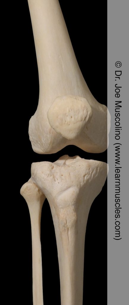
Anterior view of the knee joint on the right side of the body.
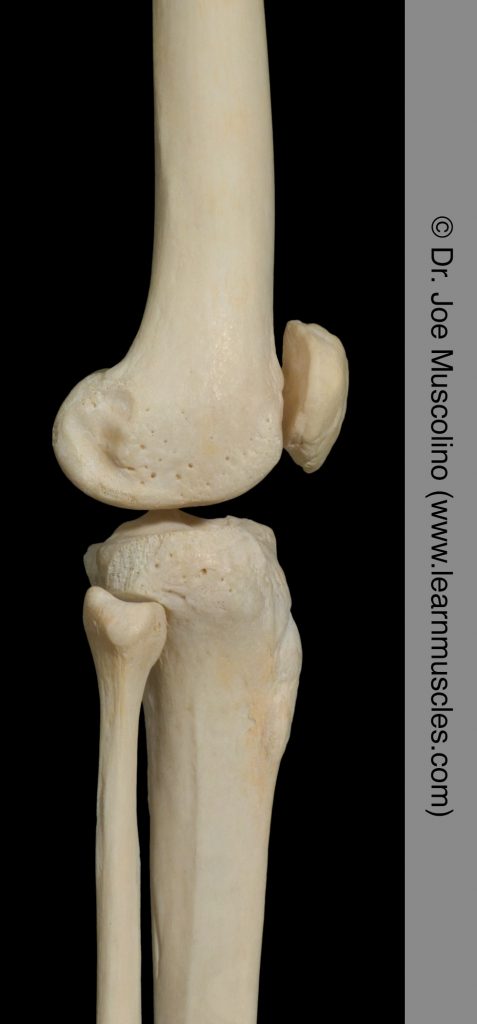
Lateral view of the knee joint on the right side of the body.