- There are five metacarpal bones in the hand.
- They are named #1, #2, #3, #4, and #5, from the lateral/thumb side to the medial/little-finger side.
- A metacarpal with its associated phalanges is called a ray.
- Metacarpal #1 is within the first ray, the ray of the thumb.
- Metacarpal #5 is within the fifth ray, the ray of the little finger.
- The metacarpals articulate with:
- the phalanges distally, forming the metacarpophalangeal joints.
- the carpals proximally, forming the carpometacarpal joints.
- each other, forming the intermetacarpal joints.
NOTES:
- Each metacarpal has an expanded proximal end called the base, and an expanded distal end called the head.
- There are two sesamoid bones located on the palmar side of the head of the first metacarpal.
- The first metacarpal (the metacarpal of the thumb) may be the shortest, but is the largest metacarpal.
- The carpometacarpal joint of the thumb is known as the saddle joint of the thumb.
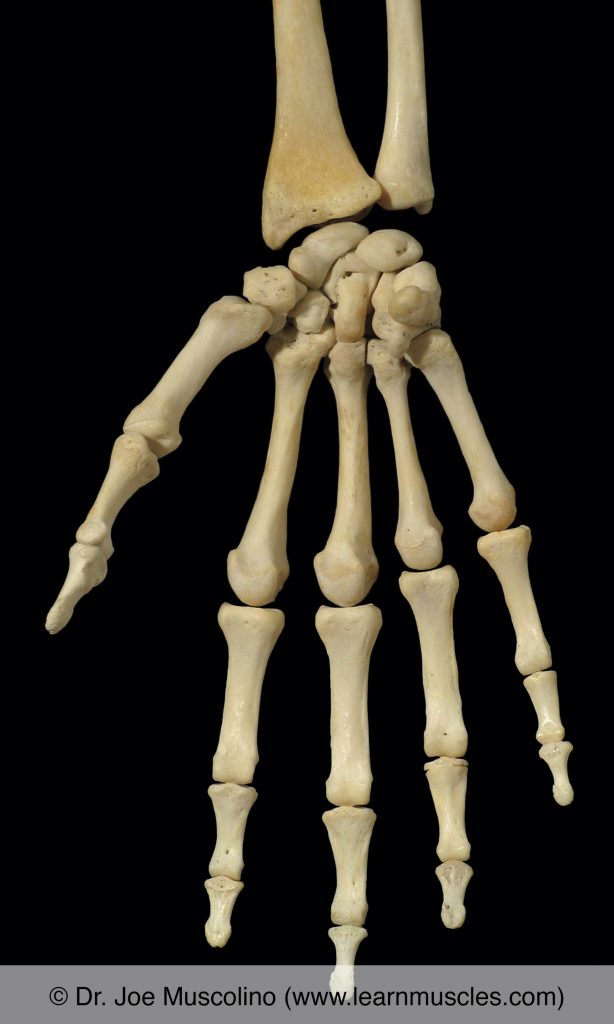
Anterior view of bones of the right hand.
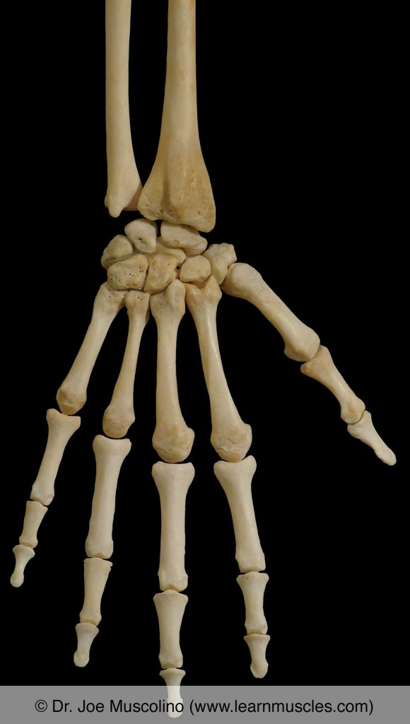
Posterior view of bones of the right hand.
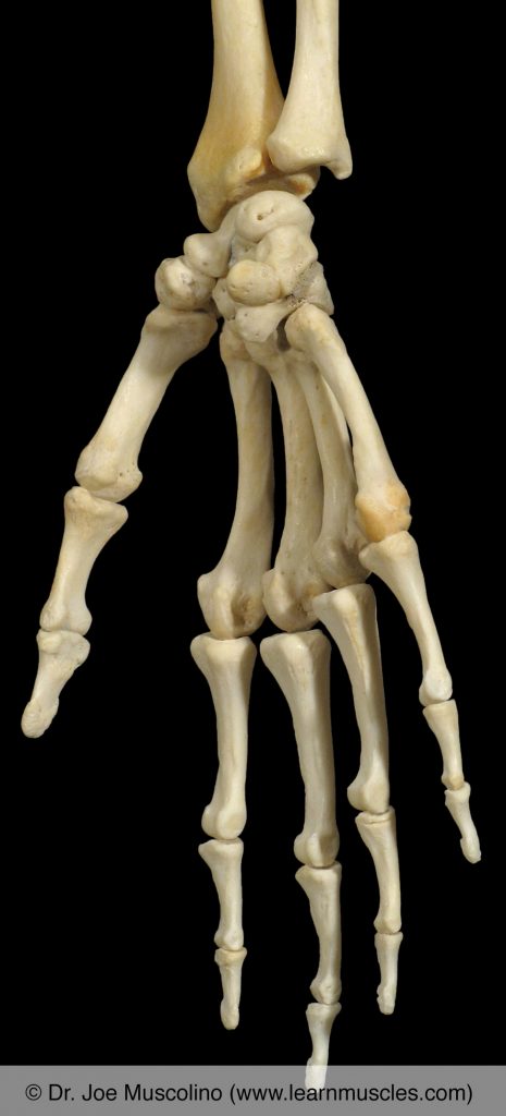
Oblique (anteromedial) view of bones of the right hand.
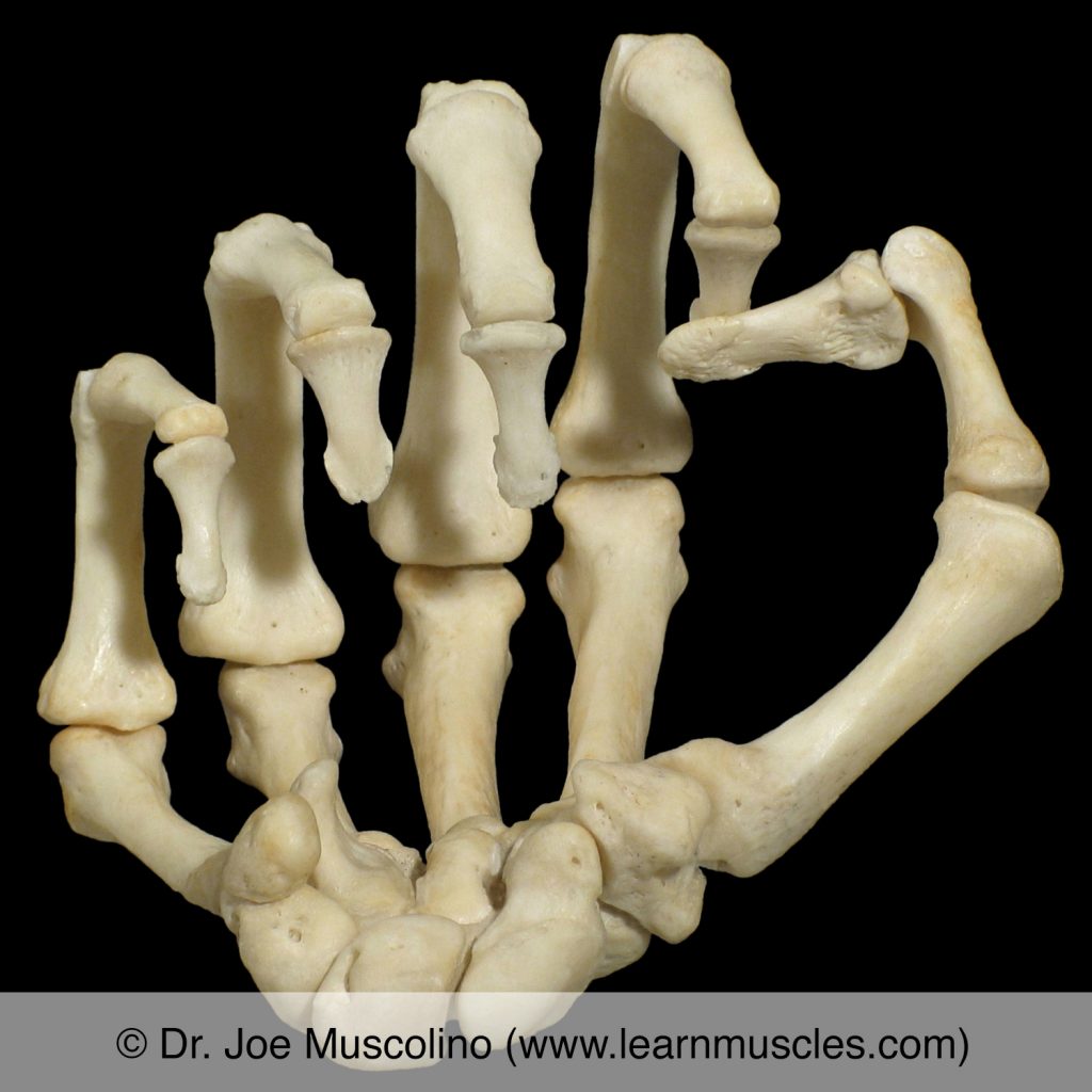
Bones of the wrist and hand in the posture of a fist on the right side.