- Click here for access to the full Anatomy Glossary.
- Right click on the image for a downloadable file of this muscle.
- Use of this artwork requires proper credit to be given (Permission: Dr. Joe Muscolino. www.learnmuscles.com – art work Giovanni Rimasti)
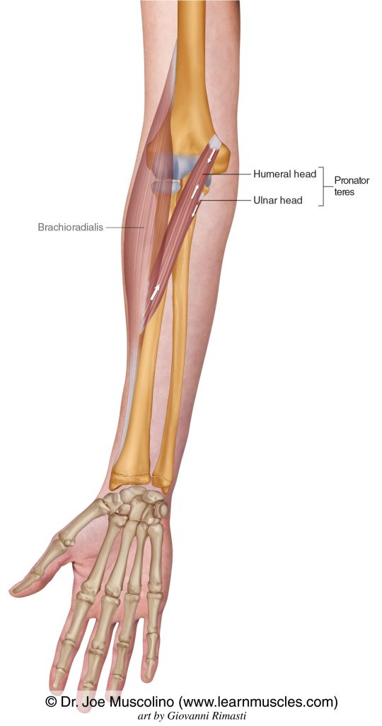
The Pronator Teres is in the Superficial Layer of the Anterior Compartment of the forearm.
The Pronator Teres has two heads: Humeral Head and Ulnar Head.
ATTACHMENTS:
ACTIONS:
- Pronates the forearm at the radioulnar joints.
- Flexes the forearm at the elbow joint.
NOTES:
- The pronator teres is one of five muscles of the common flexor belly/tendon that is involved with medial elbow tendinopathy (aka golfer’s elbow).
- The median nerve runs between the two heads of the pronator teres. If the pronator teres is tight (overly facilitated), it can compress the median nerve causing symptoms that mimic carpal tunnel syndrome. This condition is called pronator teres syndrome.