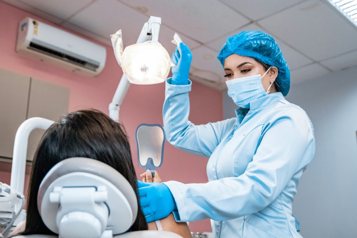
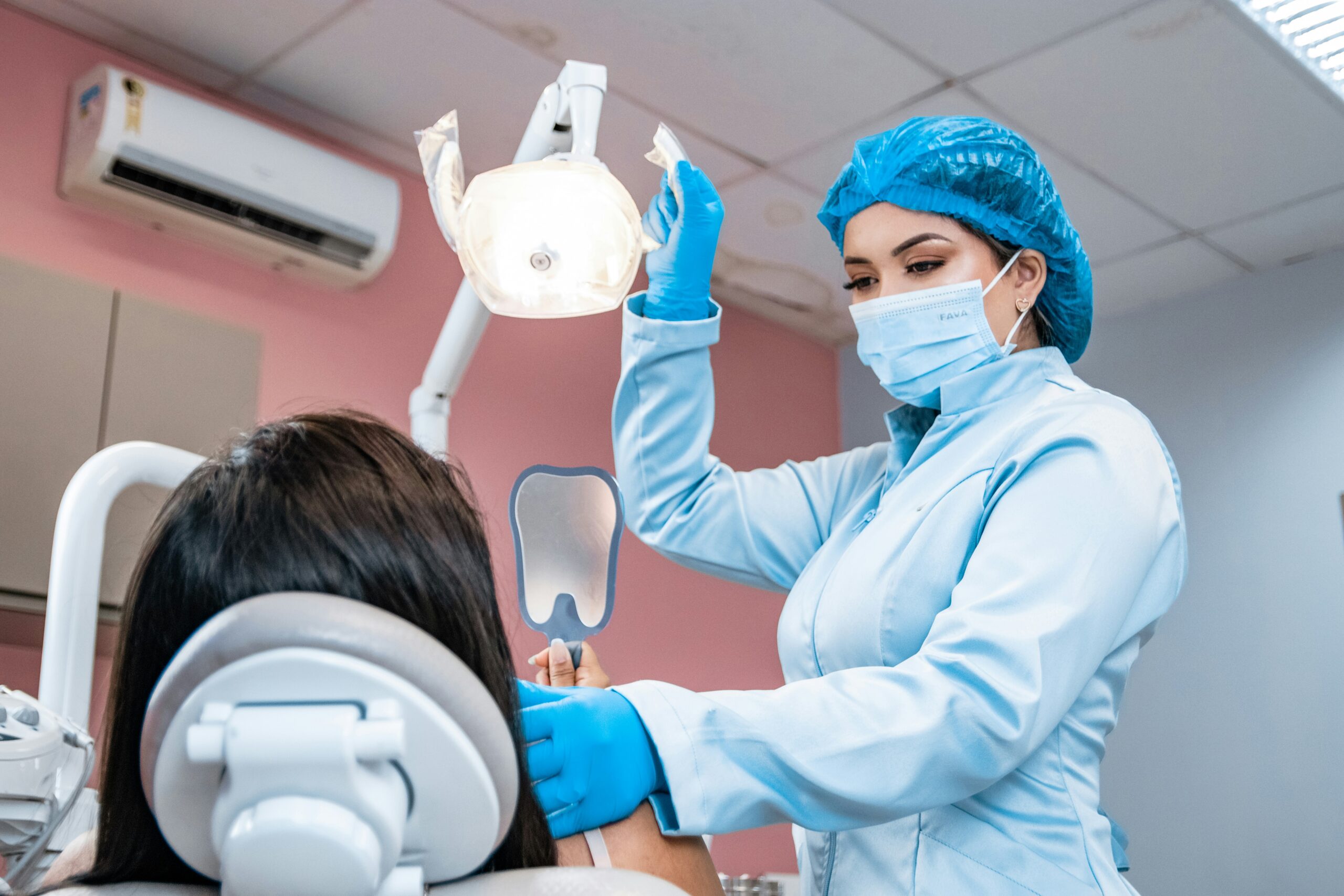
The impaction of the third molar often leads to intricate biomechanical alterations within the mouth cavity. To manage impactions an accurate knowledge of myofascial anatomy, jaw kinematics and neuromuscular responses is required. After wisdom tooth extraction, patients commonly report localized muscular tension, biting alignment alterations, and altered temporomandibular joint dynamics during the healing phase. While working with skilled professionals such as the clinical team at Smile By Design, surgical planning takes into account not only the removal of the teeth but also the long term integrity of the surrounding masticatory systems.
North York Smile Center
Neuromuscular and Myofascial Strain of Surgical Exposure
The removal of lower third molars necessitates prolonged jaw opening and the use of surgical forces near sites of significant muscle attachment. During the whole procedure the masseter, medial pterygoid and temporalis muscles are continuously isometrically stretched. Deeply impacted teeth may result in prolonged dislocation of the jaw which may stretch myofascial fibers beyond their resting length and may result in post-surgical muscle guarding for protection.
Protective muscle splinting is an instinctive neuromuscular reaction that serves to immobilize the joint and protect injured tissues. This constriction often results in transient trismus (limited jaw opening). Patients frequently describe the presence of tight knots and hyperirritable trigger points in the masseter and deep pterygoid muscles. Understanding this muscular response helps practitioners to guide patients through specific rehabilitative exercises to safely regain complete range of motion.
Temporomandibular Joint Kinematics and Ligamentous Strain
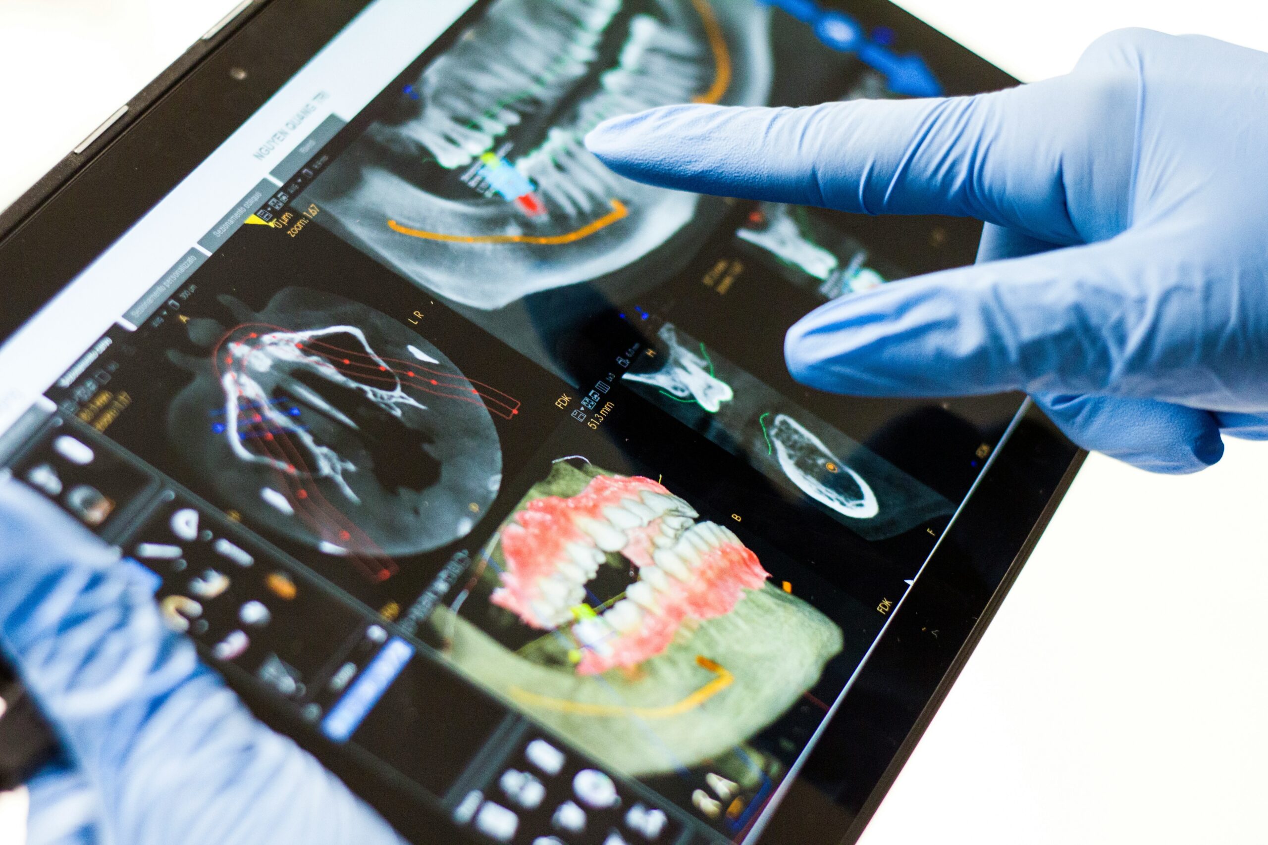
The temporomandibular joint is a delicate balance of the articular disc, condyle and surrounding capsular ligaments. Downward and backward stresses on the mandible during difficult extractions of third molars may stress the temporomandibular ligament and the sphenomandibular ligament. If the lower jaw is not adequately stabilized during the extraction, this pressure can change the relationship between the disc and the condyle.
Mechanical stress on the joint capsule can lead to localized inflammation and brief clicking or popping of the joint or dull pain near the ear. Patients with pre-existing joint hypermobility, or subclinical temporomandibular disorders, are particularly susceptible to post-extraction flare-ups. Supportive bite blocks in surgical protocols might decrease unwanted mechanical leverage on the contralateral joint capsule during molar elevation.
Secondary Muscular Compensation and Occlusal Changes
Removal of third molars modifies the distal limit of the dental arch. Removal of impacted third molars that were applying anterior pressure on the second molar changes the tactile information received by the periodontal ligament receptors. The brain responds to these slight occlusal changes by changing the firing patterns of the masticatory muscles during chewing and swallowing.
As the occlusion stabilizes subsequent muscle compensation may ensue. Uneven activity of the masseter and temporalis muscles may adapt minor alterations in bite contact. If not corrected, this unequal muscle recruitment can lead to chronic cervical spine stress, tension headaches, and myofascial pain syndrome. After surgery, mild exercises to bring the jaw back into alignment serve to re-educate the proprioceptors and reestablish balanced activation of the muscles.
Rehabilitation After Surgery of the Masticatory System
Targeted rehabilitation accelerates recovery and prevents long-term myofascial dysfunction after molar excision. After the acute inflammation has passed, patients benefit from gradual therapy programs to relax hypertonic muscles and restore full joint mobility.
Rehabilitation’s main practices include:
Moist Heat: Heat applied to the lateral jaw will relax tight masseter fibers and will enhance local blood flow to facilitate tissue recovery.
Gentle Active Range-of-Motion Drills Controlled, pain-free jaw opening and lateral deviation exercises prevent the adhesion of scar tissue within the joint capsule.
Gentle manual pressure is administered to trigger points along the masseter and medial pterygoid muscles to loosen tight muscles.
Proprioceptive Re-Education: Controlled biting exercises induce a symmetrical muscle activation on both sides of the mandibula.
Working with experienced dental surgery teams, you can be sure that hard and soft tissue structures will be properly cared for during the recovery process. By treating the anatomical effect on the jaw muscles and joints, recovery difficulties are minimized and a lasting neuromuscular balance is achieved.
Written by Milos Radakovic