
Assessment/Diagnosis of adductor strain (groin pull):
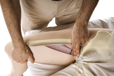
Palpation of the adductor longus. Resistance to adduction of the thigh is added with the other hand placed on the distal medial thigh. Permission: Joseph E. Muscolino. The Muscle and Bone Palpation Manual, with Trigger Points, Referral Patterns, and Stretching, 2ed. (2016), Elsevier.
Assessment/diagnosis of an adductor strain (groin pull) follows from the signs and symptoms. In the acute stage, the location of the client’s/patient’s pain will indicate the area of strain, and palpation there will further elicit pain. To determine which adductor muscle(s) is /are involved, the adductor longus’ proximal tendon can be used as a landmark for palpation because it has such a prominent proximal tendon. The pectineus lies directly lateral to it, and the gracilis lies directly medial to it. The adductor magnus is posterior to the gracilis. And the adductor brevis is usually located deep (posterior) to the longus (sometimes a small portion of the adductor brevis can be palpated between the adductor longus and the gracilis). Palpation is most successfully formed by dropping immediately off the pubic bone and then resisting the client from adducting the thigh at the hip joint against resistance and feeling for the engagement of the musculature to confirm that you are indeed on the adductor group.
During the acute phase of an adductor strain, swelling will also be palpable and possibly visible. Active adduction range of motion and manual resistance to adduction will increase the client/patient’s pain. These tests will most likely also be positive for thigh flexion (or extension if the adductor magnus is involved). Thigh abduction range of motion, whether it is active or passive, will also increase pain because it stretches the torn adductor muscle fibers. Extension range of motion might also cause pain because it stretches the more anterior fibers of the adductor musculature (flexion stretches the adductor magnus).
Once the acute stage of an adductor strain has resolved and the client’s/patient’s adductor strain is chronic, assessment is usually done by palpating for global tightness of adductor musculature and/or myofascial trigger points or fascial adhesion. Stretching the adductor musculature will also usually result in decreased range of motion into abduction, and perhaps extension or flexion depending upon whether it is anterior or posterior adductor fibers that are strained. If the adductor musculature is globally tight, postural exam might reveal genu valgus on that side (knock knees) due to the increased thigh adduction, or a high iliac crest on that side.
Differential assessment of adductor strain:
There is usually no difficulty differentially assessing an acute adductor strain from other conditions. However, a chronic adductor strain should be discerned from simple global tightness, myofascial trigger points, or fascial adhesions that exist without the provocation of an adductor strain. Given the proximity of the distal belly/tendon of the iliopsoas, it is important to be precise with palpation and differentially assess the adductors from the iliopsoas. Hip joint degenerative osteoarthritic changes also need to be differentially assessed.
