
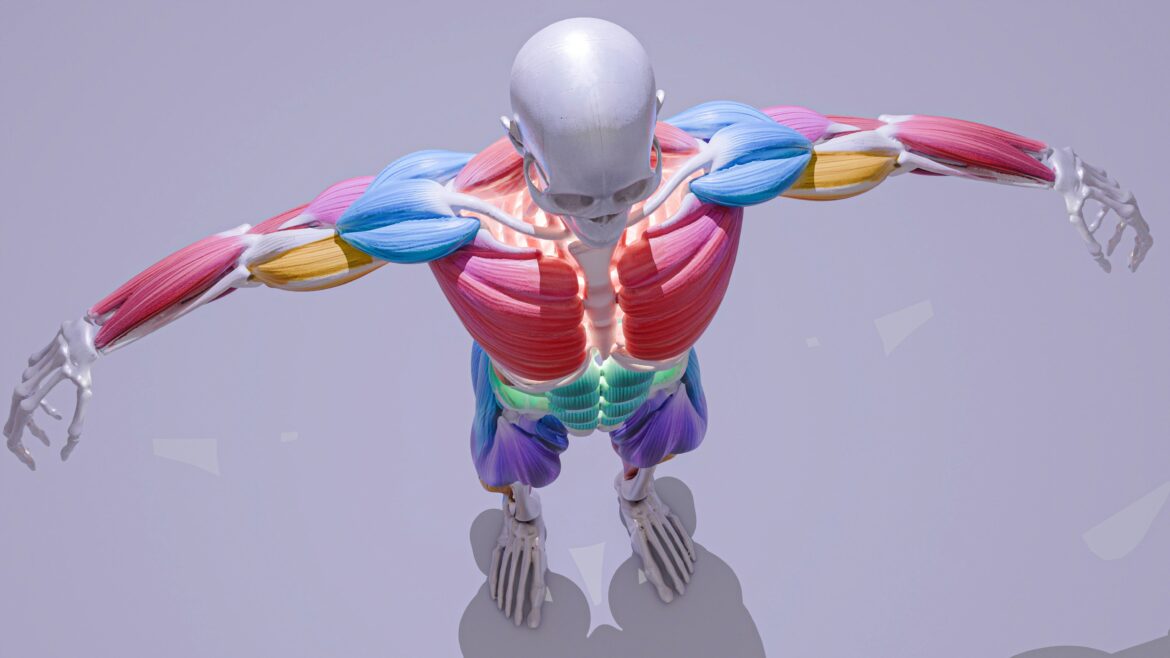
Skeletal muscles normally contract in response to signals from the central nervous system. However, high-intensity focused electromagnetic (HIFEM) technologies can initiate intense muscular contractions through externally applied stimulation. This article focuses on the physiology of muscle activation, rather than the aesthetic or cosmetic benefits often marketed alongside these devices. By generating rapidly changing magnetic fields, this technology can stimulate peripheral motor neurons without requiring a voluntary motor command from the brain. The process moves from electromagnetic energy to motor-neuron activation and finally to skeletal muscle contraction. While this mechanism induces substantial tension within the tissue, it functions differently from normal voluntary movement and cannot serve as a complete replacement for conventional exercise.
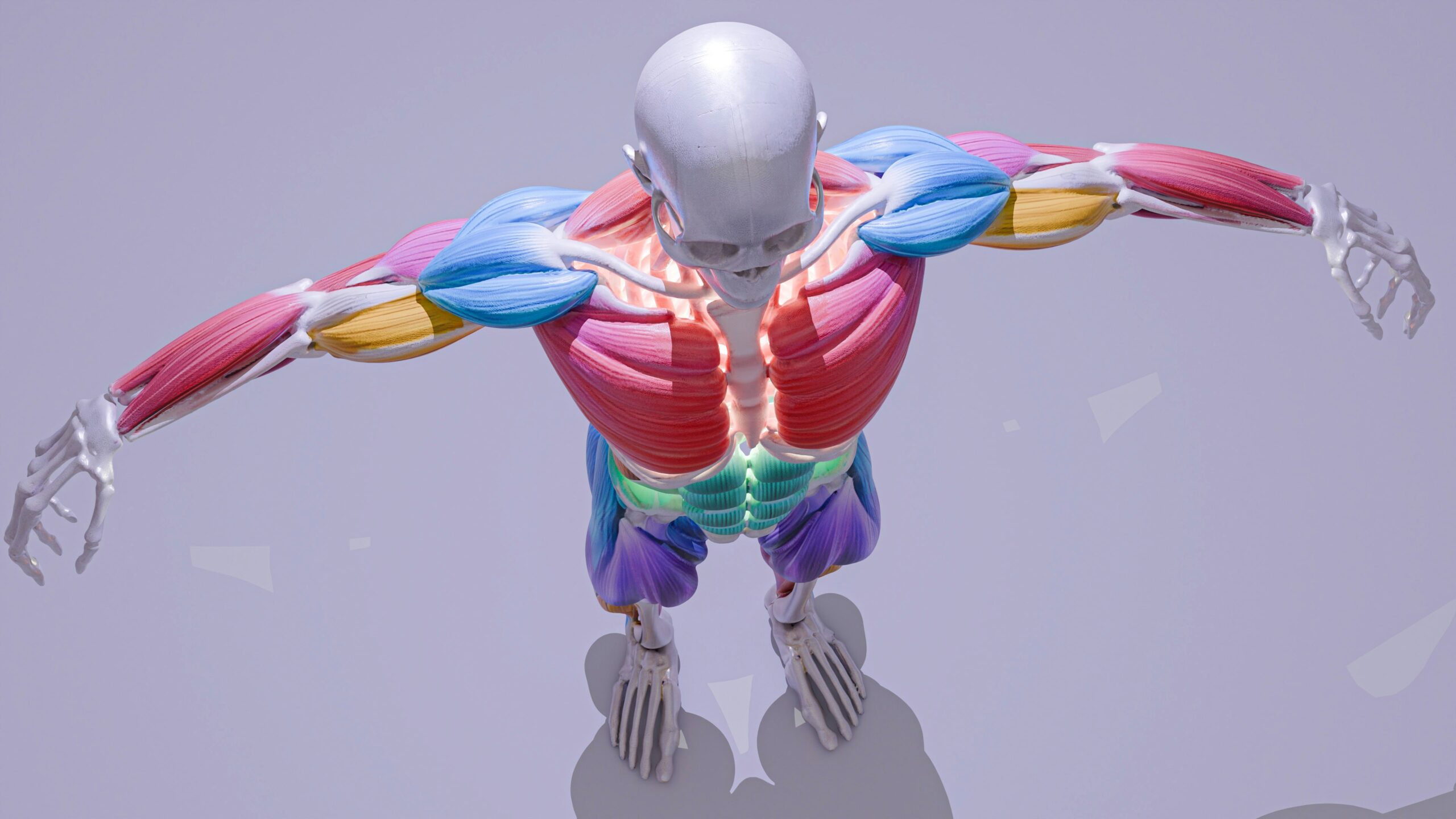
What Is Emsculpt Muscle Stimulation?
High-intensity focused electromagnetic (HIFEM) technology uses rapidly changing magnetic fields that propagate through tissue and induce electrical currents. Published descriptions of HIFEM technology report magnetic fields operating in the low-kilohertz range, with specific parameters varying by device and protocol. During a session, an applicator is positioned over a targeted skeletal muscle group. Rather than beginning with a voluntary motor command originating from the brain, the muscular contraction is externally initiated.
According to Faraday’s law of electromagnetic induction, these changing magnetic fields induce secondary electric currents within the body. The induced currents stimulate peripheral motor neurons supplying the underlying muscle fibers without requiring voluntary activation. Once those motor neurons are depolarized, they generate action potentials that travel toward the neuromuscular junction and initiate skeletal muscle contraction.
How Electromagnetic Energy Triggers a Muscle Contraction
To understand how this process causes a muscle to contract, it is necessary to trace the sequence from energy generation to cellular response. The process begins with a rapidly changing magnetic field. Published Emsculpt studies have reported field strengths up to about 1.8 Tesla, although field strength varies by platform, applicator, and treatment protocol. As these magnetic fields pass through the body, they induce secondary electrical currents in the targeted tissue through electromagnetic induction.
In HIFEM technology such as Emsculpt, the induced electrical currents stimulate peripheral motor neurons, generating action potentials that travel toward the neuromuscular junction. Readers researching Emsculpt may also encounter electromagnetic body-sculpting systems from other manufacturers; these should not automatically be assumed to be BTL EMSCULPT devices, because their hardware, treatment parameters, regulatory status, and clinical evidence may differ.
From this point, the process mirrors typical excitation-contraction coupling. The action potential travels along the sarcolemma and deep into the transverse (T) tubules, triggering the release of calcium ions from the sarcoplasmic reticulum. The calcium binds to troponin C, shifting the tropomyosin-troponin complex to expose active sites on the actin filaments. Myosin heads then bind to these exposed sites, initiating the power stroke that results in skeletal muscle contraction. In essence, while the initial trigger is an external magnetic field rather than a voluntary command from the brain, the resulting mechanical shortening of the sarcomere relies on the body’s fundamental physiological machinery.
 What Are Supramaximal Muscle Contractions?
A maximal voluntary contraction (MVC) is the greatest force that can be produced by contraction of muscles by voluntary action. A supramaximal muscle contraction, generated by external muscle stimulation by means of electromagnetic muscle stimulation, can only be generated by external action. These external actions, of high frequency and intensity, cannot be reproduced by voluntary action of the corresponding muscles.
A high frequency of stimulation is generated during sessions of electromagnetic muscle stimulation. By activating the motor neurons of the muscle under investigation, individual muscle twitches can summate to high levels of tension in the contracting muscle. This is due to the fact that there is little relaxation time between individual stimuli.
In contrast to voluntary contraction, the recruitment of motor units during muscle stimulation is external to normal voluntary movement. The large number of motor units that are stimulated to contract during muscle stimulation can contract in a pattern that is very different from voluntary contraction. This pattern is determined by a number of factors including the stimulation parameters, the anatomy of the muscle, and the design of the stimulation device.
Emsculpt Contractions vs. Voluntary Exercise
Voluntary exercise and externally applied electromagnetic stimulation rely on distinct mechanisms of action. During voluntary exercise, the motor command begins within the central nervous system. The brain recruits motor units according to physiological recruitment principles, generally engaging lower-threshold motor units before progressively recruiting higher-threshold units as force demands increase. Recruitment continually changes according to the specific movement, physical load, fatigue, and overall training demands.
With electromagnetic stimulation, muscle activation is externally induced rather than initiated through a voluntary motor command. The stimulation patterns and contraction frequencies can also differ substantially from those produced during normal exercise, including periods of sustained high-frequency contraction.
Because electromagnetic stimulation does not reproduce every physiological component of voluntary movement, it should not be considered a complete substitute for conventional exercise. It does not provide the same cardiovascular conditioning, motor coordination practice, mobility adaptations, movement-specific skill development, or broader systemic benefits associated with actively performing exercise.
How Muscle Tissue Adapts to Repeated Contractions
Repeated high-intensity contractions place substantial mechanical tension and demand on targeted muscle tissue, which may initiate adaptive responses.
Muscle Hypertrophy
Muscle hypertrophy is defined as an increase in the size and volume of existing muscle fibers. Repeated externally induced contractions may contribute to localized increases in muscle size or thickness. Studies of electromagnetic muscle stimulation have reported measurable changes in muscle thickness following treatment.
For example, some systematic reviews have reported average increases of approximately 2 to 2.2 millimeters in abdominal muscle thickness following HIFEM treatment. However, individual responses vary, and published studies differ in methodology, treatment protocol, device type, measurement techniques, and study quality.
What About Muscle Hyperplasia?
Muscle hyperplasia is defined as a biological increase in the total number of muscle fibers. Some discussions surrounding HIFEM technology reference hyperplasia based largely on findings from animal research, including porcine studies.
However, human HIFEM studies have not conclusively demonstrated meaningful skeletal muscle hyperplasia. Evidence that electromagnetic body-contouring treatments increase the actual number of muscle fibers in humans remains lacking. For this reason, muscle hypertrophy and changes in muscle thickness are better-established explanations for reported structural changes in human muscle following HIFEM treatment.
Which Muscles Can Electromagnetic Stimulation Target?
Electromagnetic stimulation has been studied across multiple anatomical regions, though efficacy depends heavily on the underlying musculature. Targeted muscles frequently include the rectus abdominis, internal and external obliques, and the larger gluteal muscles. Additional applications have been directed toward the quadriceps, hamstrings, biceps, triceps, and calf musculature.
Applicator positioning directly influences which muscle groups receive the strongest electromagnetic stimulation. The magnetic field must reach the targeted motor neurons, meaning underlying anatomy, tissue depth, body position, and specific equipment design all affect how effectively stimulation reaches a particular muscle belly.
For example, deeper muscle fibers may receive different levels of stimulation than more superficial tissue. Because muscle architecture—including pennation angle, fiber orientation, and cross-sectional area—varies significantly between regions such as the biceps and gluteal muscles, not every device or applicator is anatomically suitable for every area of the body.

Emsculpt vs. Emsculpt NEO: Does RF Change the Muscle Contraction?
The original Emsculpt system uses high-intensity focused electromagnetic (HIFEM) muscle stimulation. In contrast, Emsculpt NEO combines electromagnetic stimulation with radiofrequency (RF) energy.
HIFEM remains the direct stimulus responsible for motor-neuron depolarization and the resulting muscle contractions. The RF component primarily supplies thermal energy to the treated tissues rather than directly initiating skeletal muscle contraction. Therefore, while RF changes the overall treatment by adding controlled tissue heating, the electromagnetic component remains responsible for triggering the muscle’s neuromuscular activation.
What the Science Actually Tells Us
Electromagnetic stimulation can activate peripheral motor neurons to generate strong involuntary skeletal-muscle contractions. Repeated sessions may produce structural muscular adaptations, including measurable changes in muscle thickness reported in clinical studies.
However, individual outcomes can vary significantly. Published studies differ widely in methodology, treatment protocol, device type, sample size, measurement approach, and follow-up period. Clinical studies have reported measurable increases in muscle thickness, but study quality, potential conflicts of interest, and the clinical significance of those changes remain important limitations when interpreting the evidence.
While electromagnetic technology changes how a contraction is initiated, the resulting contraction still relies on the body’s normal neuromuscular system and skeletal-muscle machinery. HIFEM treatments can therefore create a distinctive pattern of externally induced muscle activation, but they should not be viewed as a complete replacement for voluntary exercise or its broader physiological benefits.
Written by Pam Masseria (pam@95projectsgroup.com)