- Click here for access to the full Anatomy Glossary.
- Right click on the image for a downloadable file of this muscle.
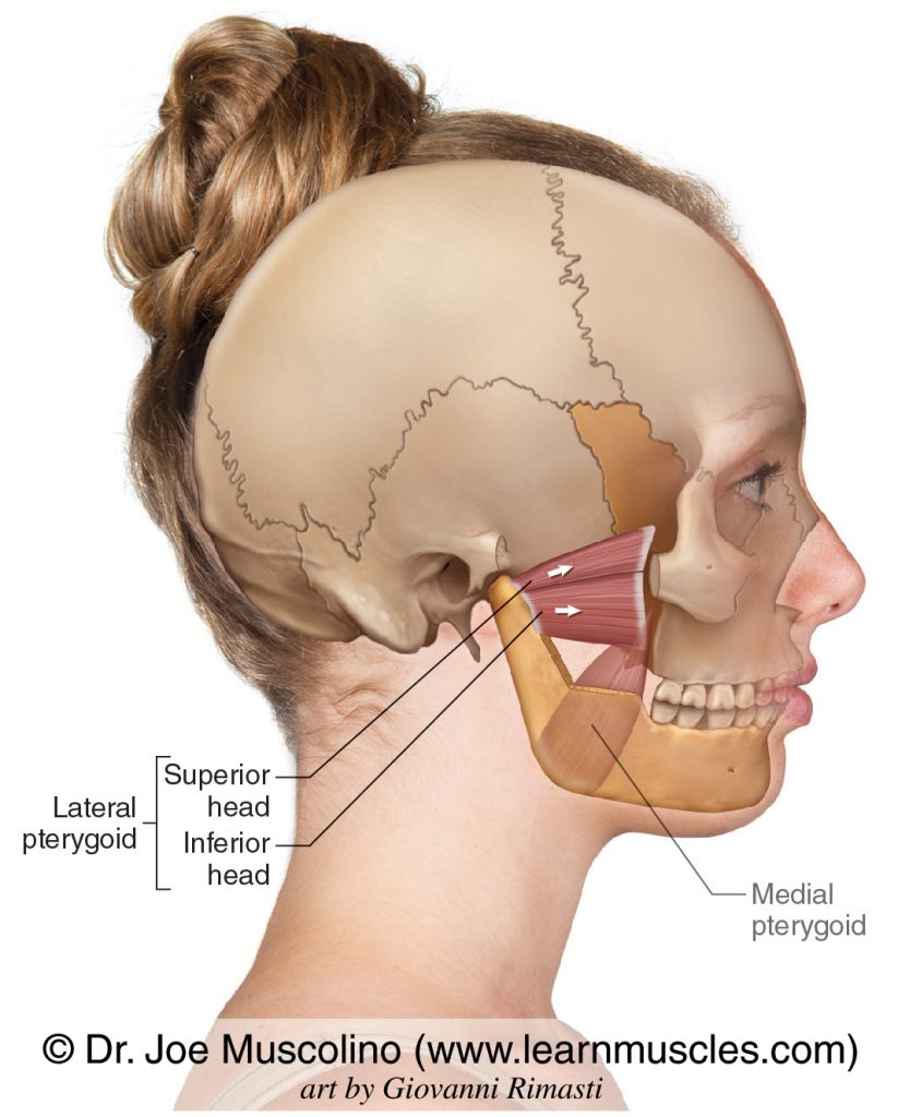
- Use of this artwork requires proper credit to be given (Permission: Dr. Joe Muscolino. www.learnmuscles.com – art work Giovanni Rimasti)
The Lateral Pterygoid is a muscle of mastication that moves the mandible at the temporomandibular joints (TMJs). The primary muscles of mastication are:
The Lateral Pterygoid has two heads: Superior Head and Inferior Head.
ATTACHMENTS:
- Sphenoid bone to the mandible and temporomandibular joint (TMJ).
ACTIONS:
NOTES:
- Having attachments directly into the articular structures of the TMJ, the lateral pterygoid is especially important to assess and treat in clients who have any sort of TMJ dysfunction.
- The lateral pterygoid is so-named because it attaches onto the sphenoid bone more laterally than the medial pterygoid.