Overview of the Joints of the Lumbar Spine and Pelvis
This blog post article is an overview of the joints of the lumbar spine and pelvis. The lumbar spine contains a disc joint and pairs facet joints. The pelvis contains the pubic symphysis and paired sacroiliac joints. For more complete coverage of the structure and function of the low back and pelvis, Kinesiology – The Skeletal System and Muscle Function, 3rd ed. (2017, Elsevier) should be consulted.
Lumbar Spinal Joints
In the lumbar spine, three joints are located between each two adjacent vertebrae: one disc joint (intervertebral disc joint) and two paired (left and right) facet joints. The disc joint is located anteriorly, and the facet joints are located posterolaterally (Fig. 7A).
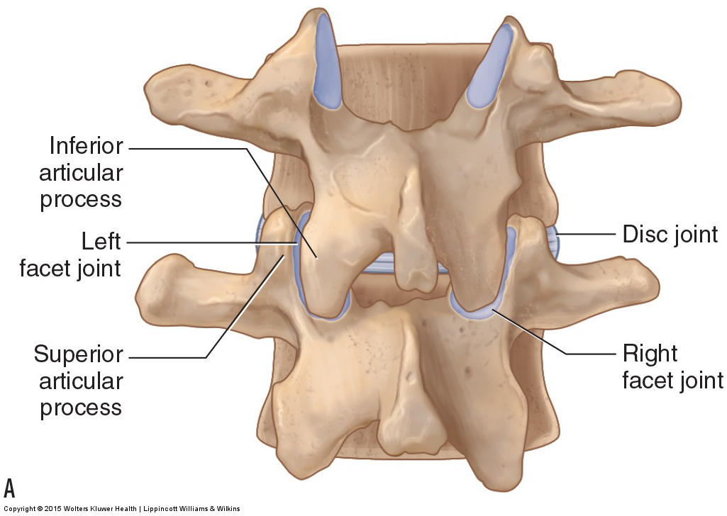
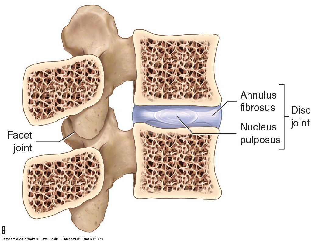
Figure 7. The disc and facet joints of the spine. (A) Posterior view. The disc joint is located anteriorly; the paired facet joints are located posterolaterally. (B) Right lateral view of a sagittal plane cross section. The disc joint is composed of the outer annulus fibrosus fibers and an inner nucleus pulposus. Courtesy Joseph E. Muscolino. Manual Therapy for the Low Back and Pelvis – A Clinical Orthopedic Approach (2015).
Disc Joint
The disc joint is a cartilaginous joint composed of outer fibers called the annulus fibrosus that encircle the inner nucleus pulposus. The annulus fibrosus is composed of 10 to 20 layers of fibrocartilaginous fibers that attach along the periphery of the bodies of the two adjacent vertebrae. The annular fibers provide a strong and stable enclosure for the nucleus pulposus.
The nucleus pulposus is a thick jellylike substance located within the disc joint (Fig. 7B). It has two main functions:
- It holds the two vertebral bodies apart, which not only creates a larger intervertebral foramen where the spinal nerve enters and exits the spine but also allows the disc joint a greater range of motion.
- It provides cushioning to the spine.
As a whole, the disc joint itself has three major functions:
- The disc joint bears the weight of the body above it. The increasing size of the vertebral bodies and discs descending the spine helps the disc joints bear the increasing weight of the body above it.
- The disc joint’s thickness allows for a great deal of motion. Overall, the intervertebral discs comprise 25% of the height of the entire spine. The greater the relative height of the discs compared to vertebral body height, the greater the possible range of motion at that region of the spine.
- The intervertebral discs help to absorb shock.
(Note: Click here for an article on pathologic disc conditions.)
Facet Joints
Each facet joint is a synovial joint that is located between the inferior vertebra’s superior articular process and the superior vertebra’s inferior articular process (see Fig. 7). It is called a facet joint because the joint surface of each of the articular processes has a facet (a smooth flat surface) on it. The scientific name for facet joints is zygapophyseal joints, hence they are also often known as Z joints. Facet joints function to guide motion at that segmental joint level of the spine. The term segmental joint level refers to a specific joint level of the spine that includes the disc and facet joints at that level. For example, the joint between L3 and L4, known as the L3-L4 joint, is a segmental level; the L4-L5 joint is another segmental level. The joint between L5 and the base of the sacrum is known as the L5-S1 joint, or simply the lumbosacral joint. An understanding of the posture of the lumbosacral joint is extremely important toward understanding pelvic tilt posture and its effect on the lumbar lordotic curve.
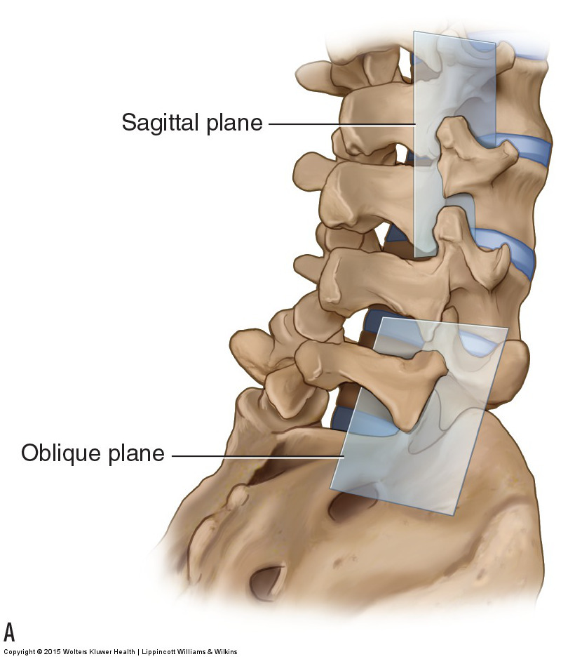
The disc joint determines how much vertebral motion is possible at a particular segmental level, and the facet joints determine the type of motion (i.e., the direction of motion) that can occur there. In the lumbar spine, the plane of the facets is vertically oriented in the sagittal plane. An exception to this is at the L5-S1 level where the facet plane is oriented in an oblique plane that is very close to the frontal plane (Fig. 8A). Note: Figure 8B is a review of the three cardinal (major) planes of the body. The three cardinal planes are the sagittal, frontal (also known as coronal), and transverse. Any plane that is not perfectly sagittal, frontal, or transverse is an oblique plane.

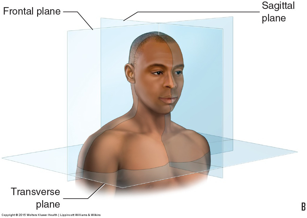
Figure 8. Plane of the facet joints of the lumbar spine. (A) The plane of the lumbar facets is oriented in the sagittal plane, except for the facet plane of the lumbosacral joint, which is oriented in an oblique plane that is close to the frontal plane. Right posterolateral oblique view. (B) Review of all three cardinal (major) planes of the body. Courtesy Joseph E. Muscolino. Manual Therapy for the Low Back and Pelvis – A Clinical Orthopedic Approach (2015).
Because of the sagittal plane orientation of the lumbar facets, the lumbar spine moves extremely well in flexion and extension (sagittal plane motions). Because the facets of the lumbosacral joint are oriented approximately within the frontal plane, right and left lateral flexion motions occur more freely at that level. It is important to be aware of the type of motion that each level of the spine allows when performing joint mobilization.
Pelvic Joints
Pelvic joints can be divided into two categories: joints between the pelvis and adjacent body parts and joints located within the pelvis. The lumbosacral joint is located between the pelvis and trunk (more specifically, between the sacrum of the pelvis and L5 of the lumbar spine) (see Fig. 2 of Bones of the Lumbar Spine and Pelvis blog post), and the hip joints are located between the pelvis and the thighs (more specifically, the femurs of the thighs) (see Fig. 6 of Bones of the Lumbar Spine and Pelvis blog post). Within the pelvis, there are two SIJs and one symphysis pubis joint. Each SIJ is located posteriorly between the sacrum and the iliac portion of the pelvic bone on that side (see Figs. 5A and 6 of Bones of the Lumbar Spine and Pelvis blog post). The symphysis pubis joint is located anteriorly between the two pubic bone portions of the pelvic bones (see Fig. 6 of Bones of the Lumbar Spine and Pelvis blog post).
Note: For an article on the relationship between the psoas major muscle and the sacroiliac joint. click here.
Note: This is the second in a series of 8 blog post articles on the anatomy and physiology of the lumbar spine and pelvis.
The blog post articles in this series are:
- Bones of the Lumbar Spine and Pelvis
- Joints of the Lumbar Spine (disc & facet) and Pelvis
- Motions of the Joints of the Lumbar Spine
- Motions of the Joints of the Pelvis
- Muscles of the Lumbar Spine
- Muscles of the Pelvis
- Ligaments of the Lumbar Spine and Pelvis
- Precautions for Manual Therapy of the Lumbar Spine and Pelvis
