
Postural dysfunction is one of the most commonly treated presentations in manual therapy, and one of the most consistently misunderstood. The standard advice to “sit up straight” treats posture as a behavioral problem, a matter of attention and effort. Clinically, that framing misses most of what is actually happening. Postural dysfunction is a neuromuscular and structural problem, and addressing it effectively requires understanding the underlying mechanisms rather than cueing the patient to fight against them.
This article examines the primary drivers of postural breakdown, how compensatory patterns develop and self-reinforce, and where spinal segmental function fits into the picture for manual therapists and movement professionals working with this population.

Postural Dysfunction Is a System Problem, Not a Habit Problem
The nervous system organizes posture around the path of least resistance. When sustained loading, repetitive movement patterns, or structural asymmetry shifts that path, the neuromuscular system adapts. Muscles that are chronically shortened or lengthened change their resting tone, alter their recruitment timing, and eventually recalibrate what the nervous system registers as neutral. The result is a posture that feels normal to the patient even when it is significantly deviated from ideal alignment.
This recalibration is the core of why postural correction is difficult. The patient is not choosing to slouch. They are expressing a pattern their nervous system has encoded as baseline. Interventions that only address the surface expression of that pattern, without addressing the underlying neuromuscular encoding, produce temporary results at best.
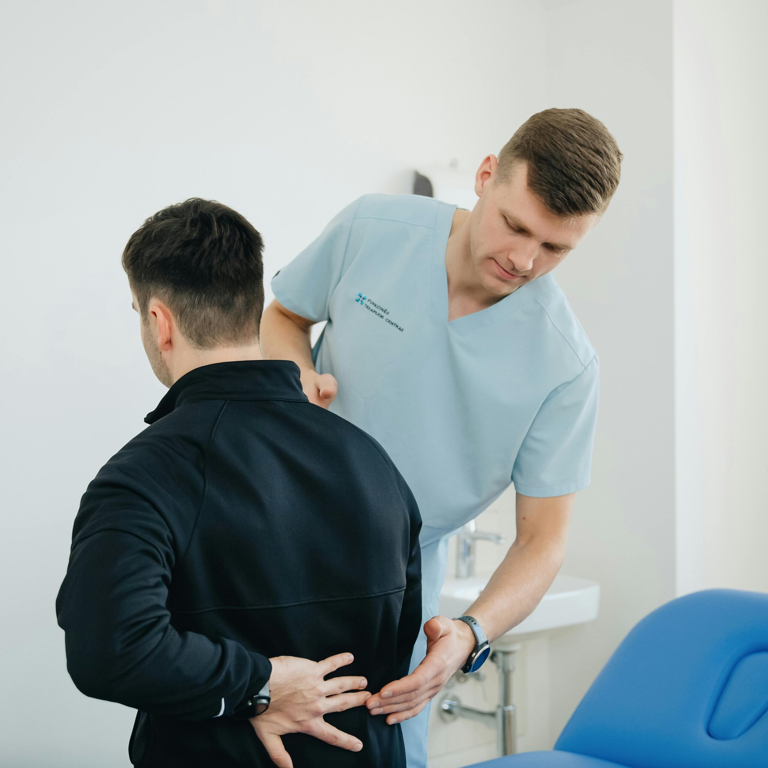
For practitioners in the San Francisco Bay Area and beyond, spinal adjustment and chiropractic care is one component of a broader intervention framework that targets segmental restriction and proprioceptive disruption at the source, rather than managing the compensatory patterns downstream.
Upper Crossed Syndrome: The Most Common Postural Pattern in Clinical Practice
Janda’s upper crossed syndrome remains the most clinically useful framework for understanding the postural dysfunction most practitioners see daily. The pattern involves predictable tightness and inhibition across two intersecting axes:
- Tight: Upper trapezius, levator scapulae, pectoralis major and minor, sternocleidomastoid
- Inhibited: Deep cervical flexors, lower and middle trapezius, serratus anterior, rhomboids
The resulting presentation includes forward head posture, increased cervical lordosis, elevated and protracted scapulae, and a rounded thoracic kyphosis. In isolation, each of these deviations creates local loading problems. In combination, they create a self-reinforcing cycle: the tight structures pull the skeleton toward the dysfunctional position, the inhibited structures lack the capacity to counteract that pull, and the nervous system continues to register the deviated position as neutral.
What makes upper crossed syndrome particularly relevant for manual therapists is that the pattern does not resolve through strengthening the inhibited muscles alone. Facilitated muscles actively suppress the recruitment of their antagonists. Until the tight structures are addressed and segmental mobility is restored, the inhibited muscles cannot be effectively recruited regardless of how diligently the patient trains them.

Lower Crossed Syndrome and the Pelvis as the Foundation
Lower crossed syndrome follows the same organizational logic. Tight hip flexors and lumbar erectors combined with inhibited gluteus maximus and abdominal stabilizers produce anterior pelvic tilt, increased lumbar lordosis, and forward displacement of the hip joint. Because the pelvis is the structural foundation for the entire spine above it, a tilted pelvis creates a cascade of compensatory adjustments through the lumbar, thoracic, and cervical regions.
A patient presenting with upper crossed syndrome features frequently has concurrent lower crossed syndrome driving the pattern from below. The forward head position that appears to be a cervical problem is, in many cases, a compensation for the altered spinal curvature that begins at the pelvis. Treating the cervical presentation without addressing the pelvic foundation produces incomplete results.
This is why a whole-spine assessment is more informative than a region-specific one for postural dysfunction. The site of pain or the most visible deviation is rarely the origin of the pattern.
The Role of Proprioception and Segmental Restriction
The mechanoreceptors in spinal joints, particularly the Type I and Type II receptors in facet joint capsules, play a central role in postural regulation. These receptors provide continuous afferent input to the cerebellum and cortex about joint position, movement velocity, and load. When a spinal segment loses normal mobility, that proprioceptive input is disrupted.
The consequences extend beyond the local segment. Altered afferent signaling from restricted joints contributes to:
- Disrupted postural reflexes and delayed neuromuscular responses
- Compensatory hypermobility at adjacent segments as the nervous system routes around the restriction
- Altered muscle recruitment patterns in the segments above and below the restriction
- Reduced accuracy of the nervous system’s internal model of spinal position
This is the clinical rationale for addressing segmental restriction as part of postural rehabilitation rather than relying solely on soft tissue work and exercise. A restricted segment is not just a local mobility problem; it is a source of degraded proprioceptive input that affects how the entire system organizes movement and posture.
Fascial Continuity and Tensegrity in Postural Patterns
Thomas Myers’ myofascial meridians provide a useful complementary framework for understanding how postural patterns propagate beyond the local muscle groups identified in Janda’s crossed syndrome models. The superficial front line, superficial back line, and spiral line in particular explain how tension or restriction at one region creates predictable loading changes at distant sites.
A shortened superficial front line, for example, contributes to the forward head and rounded shoulder pattern of upper crossed syndrome, but its influence extends inferiorly through the rectus abdominis and hip flexors to the anterior tibialis, affecting ankle dorsiflexion and foot mechanics. A practitioner assessing only the cervical and thoracic regions will miss the distal contributions to the pattern.
For manual therapists, this framework supports a more systemic approach to postural assessment: following lines of tension rather than treating isolated regions, and considering the foot, ankle, and lower extremity as potential contributors to a presentation that shows up primarily in the trunk and cervical spine.
Implications for Treatment Sequencing
Understanding the mechanisms of postural dysfunction has direct implications for how treatment is sequenced. Effective postural rehabilitation generally follows this logic:
Address segmental restriction first. Joint mobility work, whether through manipulation, mobilization, or instrument-assisted techniques, restores normal afferent signaling and creates the neurological conditions under which soft tissue and exercise interventions can be more effective.
Release facilitated muscles before strengthening inhibited ones. Attempting to strengthen inhibited muscles while their antagonists remain facilitated produces limited results. Soft tissue work targeting the tight structures, followed by neuromuscular re-education of the inhibited ones, is more productive than loading the inhibited muscles directly.
Integrate movement pattern retraining. Once mobility is restored and muscle balance is improved, the nervous system needs repetition in the corrected pattern to update its baseline. Motor control exercises, particularly those that challenge postural stability in functional positions, accelerate that recalibration.
Address contributing factors at all relevant regions. Pelvis, thorax, cervical spine, and lower extremity all contribute to the overall postural pattern. A region-specific approach that ignores the structural relationships between segments will produce improvements that are partial and often temporary.
Posture as an Expression of Load History
The clinical takeaway is that posture is not a character trait or a habit in the behavioral sense. It is the nervous system’s best current solution to the mechanical demands it has been given over time. Changing it requires changing the inputs: restoring joint mobility, rebalancing soft tissue tension, correcting movement patterns, and giving the nervous system enough repetition in the new pattern to recalibrate what it registers as neutral.
For manual therapists, that means posture is always a starting point for assessment rather than an endpoint. The visible pattern is the output. The work is understanding and addressing what is generating it.
Written by shahidshahzad26@gmail.com
