
Note: This is the seventh blog post article in a series of 11 articles on Psoas Major Function. See below for the other articles in this series on psoas major function.
We will now examine the role of the psoas major regarding motion of the spine in the sagittal plane. This is by far the most controversial aspect of psoas major functioning. Within the sagittal plane, the question is whether the psoas major creates flexion or extension of the lumbar spine.
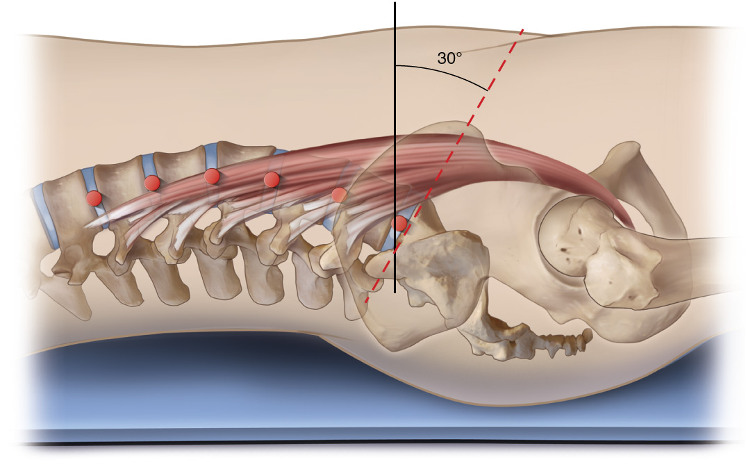
Figure 12. Right lateral view of the psoas major with a neutral pelvis/spine posture. The axes of motion at the joints are represented by red dots. Reproduced with kind permission from Joseph E. Muscolino. Art work by Giovanni Rimasti.
Overview of the Controversy
Of all of the psoas major’s functions, its effect upon the lumbar spine in the sagittal plane is by far the most controversial. Evaluating its pull on the lumbar spine in the sagittal plane is complicated by the fact that there is a different (mediolateral) axis of motion for each of the lumbar spinal joints. It is further complicated by the fact that the lumbar spine curves within the sagittal plane; and whether the lumbar spine has a normal lordotic curve, a decreased (hypolordotic) curve, or an increased (hyperlordotic) curve, can change the muscle’s line of pull relative to each of these axes. The psoas major can directly affect the degree of the lumbar curve because it crosses these joints; and it can indirectly affect the lumbar curve by changing the posture of pelvic tilt across the hip joint. And, certainly, the degree of lordotic curve is affected by whether or not the person is in anatomic position or their spine is first flexed or extended. As a result, the action of the psoas major at one lumbar spinal joint might be different than its action at another lumbar spinal joint; and each of these actions might change as the position of the lumbar spine changes. The psoas major is also a large muscle that can be considered to have upper and lower fibers, as well as anterior and posterior fibers. Consequently, many sources divide the psoas major into upper and lower parts; and others divide it into anterior and posterior parts.
Flexion versus Extension of the Spine
Scanning the literature, the controversy is immediately apparent. Numerous sources describe sagittal plane spinal function of the psoas major by stating that it can either “flex,” “bend,” “pull the trunk anteriorly,” or “raise it from a supine position” (3, 4, 6, 7, 9, 15, 16, 19, 20, 24, 27, 29, 30). But in many of these cases, whether flexion of the “trunk” refers to flexion of the lumbar spinal joints or instead refers to anterior tilt of the pelvis at the hip joints (with the lumbar spine moving along with the pelvis) is not clear because some references state that it flexes/bends/pulls the trunk anteriorly, but they also state that it increases lordosis (6, 7, 9). This might seem somewhat contradictory because flexion of the lumbar spine essentially decreases lordosis because lordosis is a curve of extension (14, 27). Other sources state that the psoas major can extend the lumbar spine (2, 4, 19, 20, 23, 25, 27). How to reconcile these results?
Effectively, we need to return to the fundamental understanding of how a muscle functions: it creates a line of pull relative to the axis of a joint. If the psoas major has a line of pull that is anterior to a lumbar spinal joint, that line of pull will cause flexion at that joint; if the psoas major has a line of pull that is posterior to a lumbar spinal joint, that line of pull will cause extension at that joint. So let’s examine the line of pull of the psoas major at the lumbar spine, or stated more accurately, let’s examine the psoas major’s lines of pull relative to the multiple lumbar spinal joints.
Upper versus Lower Fibers of the Psoas Major
Figure 12 (above) shows a lateral view of the psoas major and lumbar spine. Although there is no exact definition, a “neutral pelvis and spine” posture in the sagittal plane when in anatomic position occurs when the sacral base angle is approximately 30 degrees (14, 19). The sacral base angle is determined by measuring the angle formed by the intersection of a line drawn along the top of the sacral base and a horizontal line. In neutral position spine, we can see that the psoas major passes anterior to some of the axes and posterior to others; therefore, the psoas major can create both flexion and extension of the lumbar spine. Looking more closely, we see that, on the whole, it passes anterior to the lower lumbar spinal joints and posterior to the upper lumbar spinal joints. For this reason, many sources state that the psoas major flexes the lower lumbar spine and extends the upper lumbar spine (2, 9, 19, 20, 24, 25, 25). Tom Myers states the opposite: that the psoas major extends the lower lumbar spine and flexes the upper lumbar spine. However, he adds that this is clinical speculation and not backed up by evaluation of the mechanical axes (16, 17).
Myers does add a fascinating explanation for why the psoas major is so capable of having differing lines of pull for its upper versus lower fibers. He notes that the psoas major is actually a triangular muscle, not a fusiform muscle as it appears at first glance (17). If a muscle is triangular in shape, the fibers are not parallel; instead they have different directions and therefore differing lines of pull. Figure 13 demonstrates the psoas major of a quadruped in which we can clearly see the triangular shape. An appreciation of this triangular shape was lost when we stood up and became bipedal because the more superficial, longer, upper fibers now cover over the deeper, shorter, lower fibers. It is worth pointing out that in a quadruped, not only is the triangular shape readily apparent, but the moment arm leverage for the psoas major is much greater than in our bipedal stance.
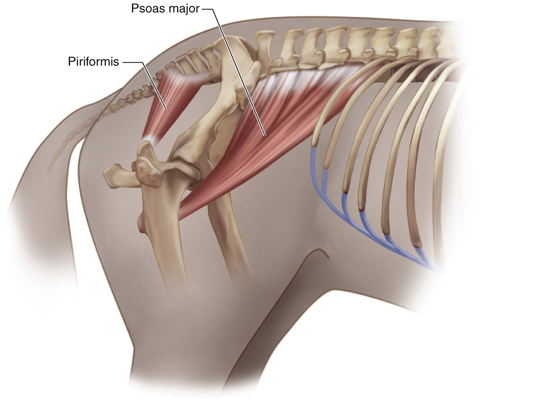
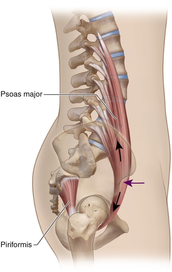 Figure 13. The triangular shape of the psoas major is much more apparent in a quadruped (A) than in a person (B, biped). Reproduced with kind permission Art work by Giovanni Rimasti. from Joseph E. Muscolino. Modeled from Myers, T. (1998). Poise: Psoas-Piriformis Balance. Massage Magazine, March/April, Figure 5B, page 77.
Altering the Lordotic Curve
It must be kept in mind that Figure 12 showed a neutral spine in anatomic position. But what happens to psoas major function if we alter the degree of lordosis? Figure 14A shows a decreased sacral base angle with a corresponding decreased lordotic curve; Figure 14B shows an increased sacral base angle with a corresponding increased lordotic curve. Comparing Figures 14A and B with Figure 12, we notice that as the lumbar lordosis decreases, the over all line of pull of the psoas major moves farther anteriorly relative to the axes; and as the lumbar lordosis increases, the over all line of pull of the psoas major moves farther posteriorly relative to the axes. This means that the psoas major flexion capability increases (and its extension capability decreases) as the curve of the spine decreases; and its extension capability increases (and its flexion capability decreases) as the curve of the spine increases. For this reason it is not simple enough to just look at a muscle when the body is in anatomic position. Muscle actions often change as we change the angles of our joints. Indeed, many sources discuss the psoas major’s variable ability to flex or extend the spine based on the degree of lordosis (10, 23, 25, 27). Ironically, the degree of lordosis itself is based on the sacral base angle, and all hip flexors, including the psoas major, if tight, will increase anterior pelvic tilt and therefore increase lumbar lordosis (4, 8, 14, 19). Therefore, the psoas major affects the lumbar spine both directly by crossing its joints, and indirectly by affecting the posture of the pelvis.
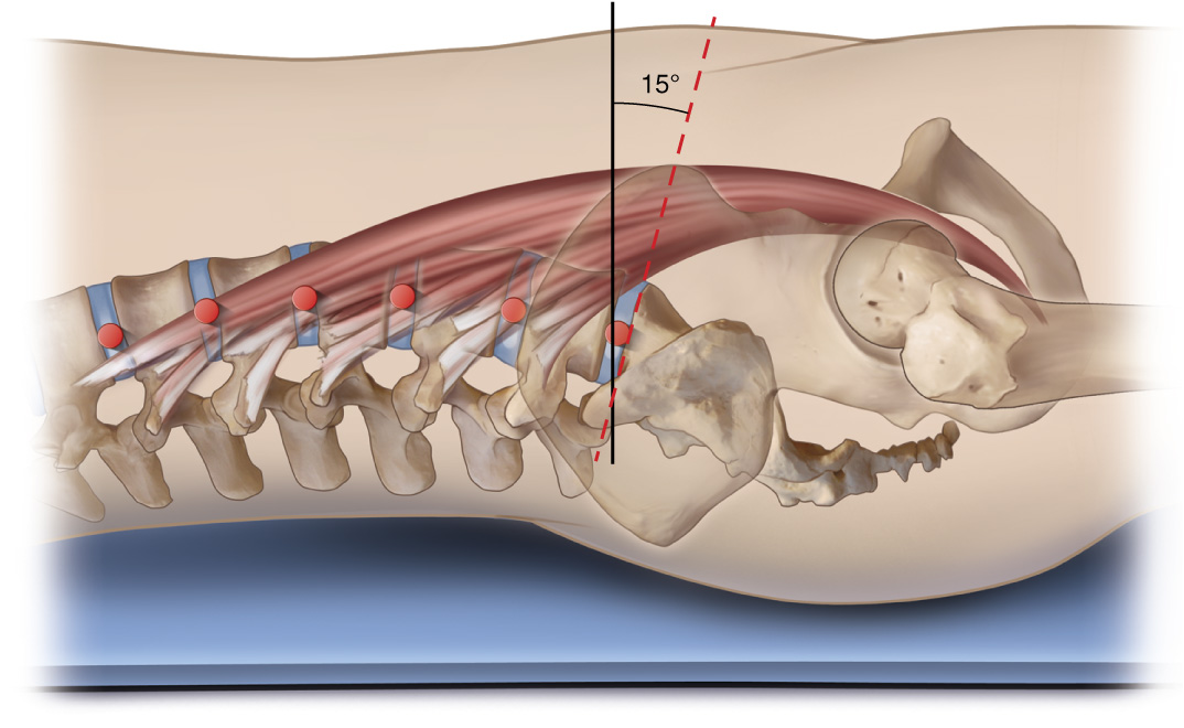
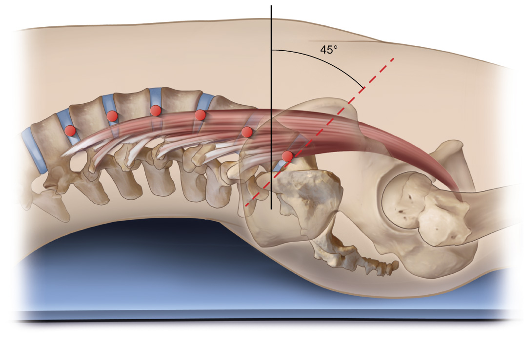
Figure 14. Lateral views of the psoas major with various degrees of lumbar lordosis. A, hypolordotic curve. B, Hyperlordotic curve. The axes at the joints are represented by red dots. Note the change in lines of pull of the psoas major as the degree of lordosis changes. Courtesy Joseph E. Muscolino. Art work by Giovanni Rimasti.
Figure 15 depicts scenario in which the person’s spine is not in anatomic position. In this scenario, the person is doing a curl-up. Similar to Figure 14A, we see that the flexion capability of the psoas major increases compared to anatomic position as the person curls upward. There are many sources that describe varying sagittal plane capability of the psoas major depending on the position of the body or the activity in which it is being engaged (6, 9, 20, 27). Engagement of the psoas major during sit-ups, crunches, and curl-ups has been especially well studied and documented by many sources (4, 15, 19, 20, 27).
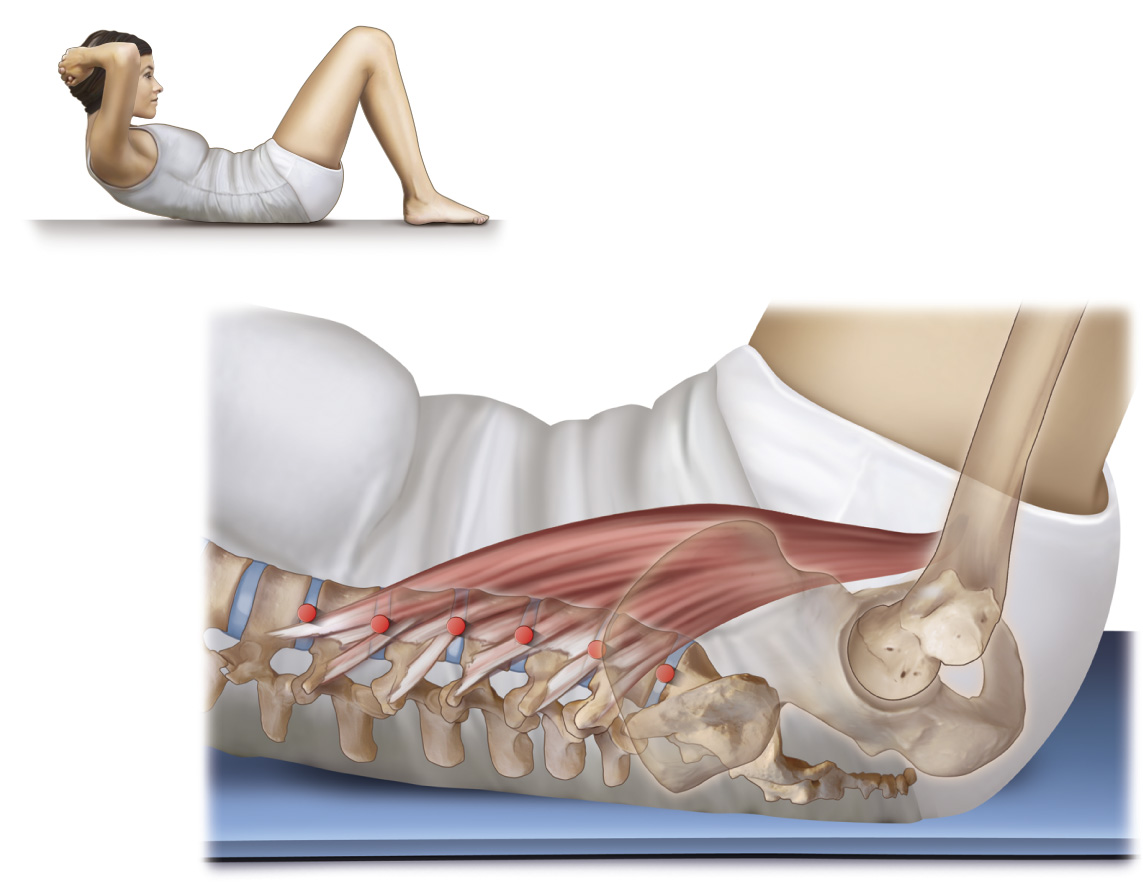
Figure 15. Lateral view of the psoas major as a person performs a curl-up exercise. Courtesy Joseph E. Muscolino. Art work by Giovanni Rimasti.
Anterior versus Posterior Fibers
There is another distinction regarding the psoas major that can be made. Not only can we look at the psoas major as having upper and lower parts, we can also divide it into anterior and posterior parts. The vertebral body and disc attachments comprise the anterior part; the transverse process attachments comprise the posterior part. Figure 16 separates the anterior versus posterior fibers at the L3-L4 level in relation to the axis of motion at the L4-L5 joint level. We can see that the anterior fibers bias toward crossing the joint anteriorly and would therefore create flexion; and the posterior fibers bias toward crossing the joint posteriorly and would therefore create extension. For this reason, many sources feel that dividing the psoas major anteriorly/posteriorly is valid (2, 7, 24). Indeed, Gibbons reports that in “…a cadaver dissection study of 24 cadavers, all specimens had a separate nerve supply for the anterior and posterior fasciculi…” He then goes on to state: “In light of this, PM (psoas major) should be considered as two distinct parts: anterior and posterior.” (2).
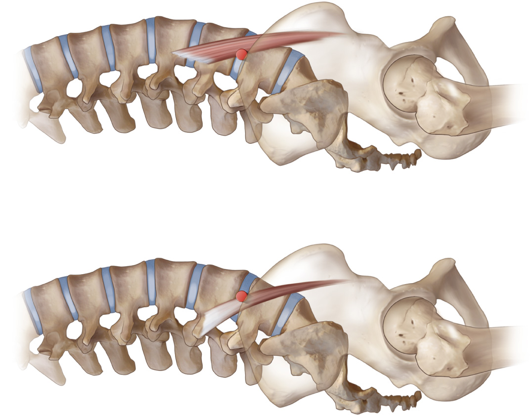
Figure 16. Lateral view of the psoas major A, Anterior fibers. B, Posterior fibers. Courtesy Joseph E. Muscolino. Art work by Giovanni Rimasti.
The Psoas Paradox
So far, analyzing the effect of the psoas major on the lumbar spinal joints has been very straight-forward and direct: compare the line of pull relative to the axis at each joint level and we will have the action of the psoas major at that joint. Unfortunately, this approach might be overly simplistic because the effect of the psoas major cannot necessarily be isolated locally. What occurs at one lumbar spinal joint level might have an effect on nearby joint levels. Looking at the body from this perspective might explain some of the seemingly contradictory effects of the psoas major upon the spine. If we look at the pull of the psoas major at the lower lumbar spinal joints, we see that it crosses anteriorly and therefore should create spinal flexion at the lower lumbar region (see Figure 12). This would imbalance the center of weight of the body anteriorly. If the nervous system wants to maintain the center of weight of the upper trunk, neck and head balanced over the pelvis, for example so that the eyes and inner ears are level to perceive the world (this is known as the “righting reflex”), it would order other musculature to compensate by creating extension of the upper lumbar region so that the center of weight of the body is brought back posteriorly to be balanced over the pelvis. Therefore, even without a strong ability of the psoas major itself to create spinal extension, and Travell and Simons report that the psoas major’s contribution toward extension is extremely weak (27), the response of the body might be to engage other musculature to extend the upper lumbar spine.
This compensation is known as the “psoas paradox” or referred to as “paradoxical lumbar lordosis (lumbar extension)” and has been cited by numerous sources as a sagittal plane effect of the psoas major upon the spine (9, 11, 15, 20, 27). Oatis makes the point though that this requires the person to have a flexible upper lumbar spine that can move into extension. Otherwise, the person might end up with a decreased lumbar lordosis and a forward lean to their posture, which would be yet another possible effect of the psoas major (20). The psoas paradox helps to resolve much of the controversy over the psoas major’s effect upon the spine and emphasizes the importance of looking at the bigger picture of muscle coordination patterns body-wide.
Click here for a list of the cited references.
Note: This is the seventh blog post article in a series of 11 articles on Psoas Major Function.
The 11 articles in the series are:
- Introduction & Muscle Biomechanics
- Biomechanics of the Psoas Major (Overview)
- Psoas Major Hip Joint Actions – Sagittal Plane
- Psoas Major Hip Joint Actions – Frontal Plane
- Psoas Major Hip Joint Actions – Transverse Plane
- Psoas Major Spinal Joint Actions – Frontal and Transverse Planes
- Psoas Major Spinal Joint Actions – Sagittal Plane
- Stabilization of the Spine by the Psoas Major
- Psoas Major and the Sacroiliac Joint
- Psoas Major and Fascial Pulls
- Summary of Psoas Major Function & Further Research
- (References)
Note: This article is modified from an article originally published in the massage therapy journal (mtj): Psoas Major Function: A Biomechanical Examination of the Psoas Major. Spring 2013 issue.
