
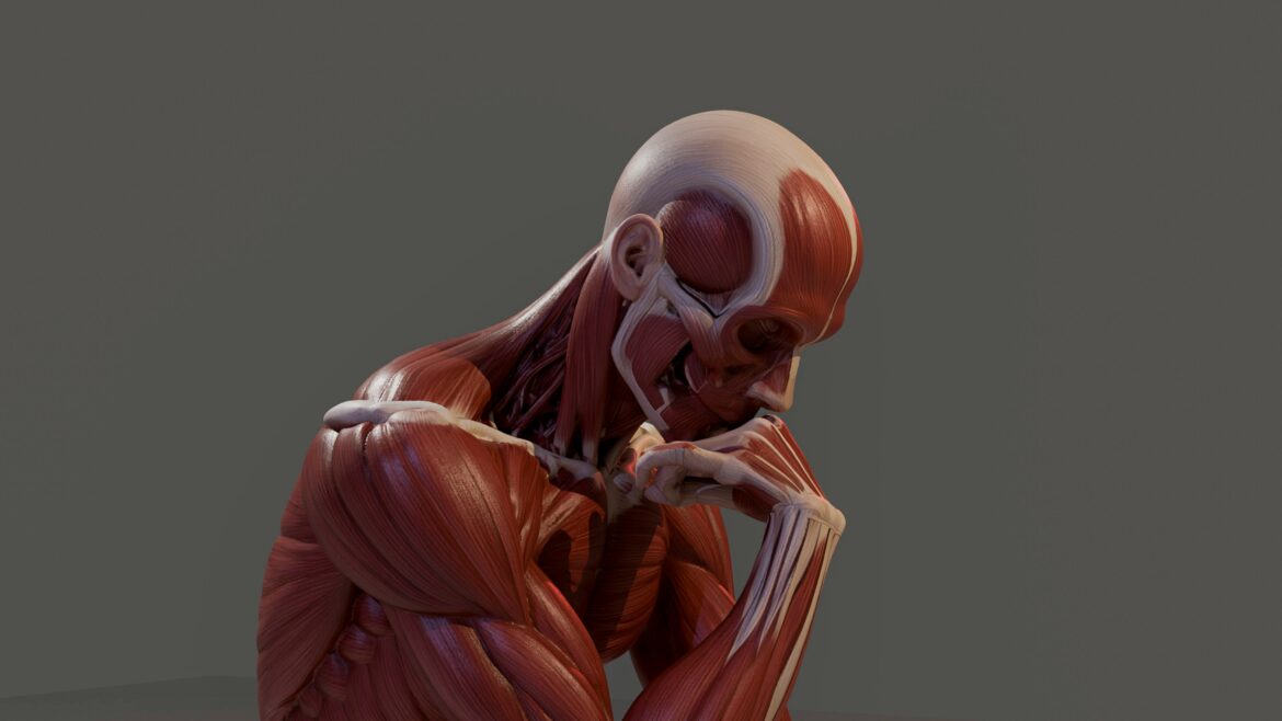
The traditional musculoskeletal model describes the events of movement. The levers, pulleys, and joints move as the muscles attach to the bones and contract. However, the living body is far more responsive than a simple diagram would suggest. All of the structures of the body, including the muscles, tendons, ligaments, and bones are subject to forces that affect them in different ways.
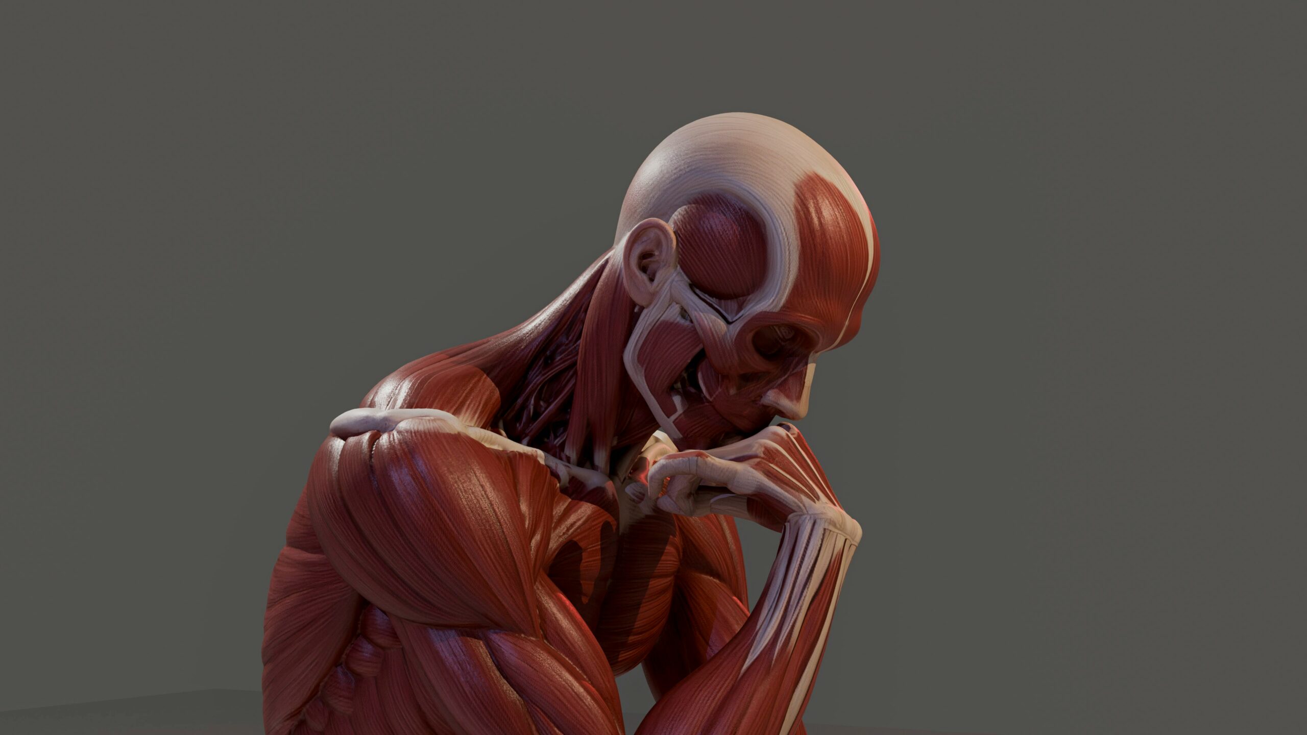
The neuro-myo-fascio-skeletal system is a dynamic system, which continuously receives and organizes information to allow optimal movement to occur. The information for movement is received from sensory receptors in the muscles, tendons, ligaments, and joints as well as from proprioceptors (sensory receptors that provide information regarding position, movement, tension, and weight of body parts). The information received by the sensory receptors is organized and movement produced.
This understanding of how tissues change shape under force, is something that can be measured and studied in detail. Therefore, when talking about anatomy, physiology and kinesiology, it is essential to remember that all of these subjects are about human movement. Therefore, the study of human movement is not just about levers and pulleys etc., but about forces acting on tissues, which then change shape.
A fundamental application of the concept of mechanotransduction for manual and movement professionals is that of a bridge between the study of anatomy and of physiology or between the study of structure and of function.
Load, Tension, Compression, and Shear
There are other forces at work while a person is moving. These forces may be quieter than others and affect various tissues. Such as the compression of a joint surface, the shear between fascial layers, the tensile load on a tendon as a limb slows down.
Kinesiology, the study of human movement, typically describes the movement of joints in relation to the actions of the attached muscles. The biceps brachii muscle for example flexes the elbow joint. The gluteus medius muscle on the other hand is involved in controlling the frontal-plane movement of the pelvis. Deep spinal stabilizers are involved in controlling the segmental movement of the spine. Other forces are also at work during movement. By studying these additional forces a more complete understanding of human movement can be gained.
- Muscle: shorten, lengthen or contract.
- Tendons: pull from the muscle to the bone.
- Fascia: distribute forces in a region rather than having them concentrated in one single structure.
- Joints: distribute compression within the surfaces of the joint and allow for glide within the joint.
- Joint Capsules: consist of soft tissues and hold the joint surfaces together, as well as the surrounding soft tissues.
The nervous system organizes and monitors the above-mentioned structures in order to control and coordinate movement.
Joints can move in less available ranges of motion. Muscles can be asked to carry loads that the muscles are unable to coordinate properly. Tissues, such as fascia, can lose their ability to adapt and therefore distribute forces less efficiently. Tissues surrounding the affected region can compensate in a number of ways by increasing range of motion, developing stiffness, requiring increased amounts of force to move, etc. in order to allow for movement in the affected region(s).
In order for a cell to respond to any given stimuli, it must first sense a change in the mechanical environment. This change in the mechanical environment is determined by the type of force that is placed upon the given tissue and/or organ. In addition, the direction in which this force is placed, the duration of time in which the force is placed upon the tissue/organ, as well as the number of times in which the force is reapplied, all play a role in determining the information that is conveyed to the neuro-myo-fascio-skeletal system.
Fascia and the Transmission of Force
Fascia gives the neuro-myo-fascio-skeletal system continuity. It surrounds muscles, blends with tendons and aponeuroses, reinforces joint capsules, and helps organize relationships between structures that are often taught separately. A muscle may have a clear origin, insertion, and action in an anatomy chart, but in the body, its force rarely stays within one isolated line.
When a muscle contracts, some of its force travels through tendon to bone, creating joint motion or stabilization. Some of that force also disperses through fascial connections into neighboring tissues. This is why movement professionals often think in terms of regions and chains instead of single muscles acting alone. Hip extension, spinal rotation, shoulder elevation, and gait all depend on coordinated tension across multiple tissues.
Epimuscular myofascial force transmission helps explain why force produced by one muscle can influence neighboring muscles and surrounding connective tissues. Rather than treating muscle action as a closed event within one muscle belly, this concept points to a wider mechanical relationship among muscles, fascia, tendons, and joints.
That relationship matters in pathomechanics. If force transmission becomes less balanced, one area may move too much while another becomes relatively restricted. A joint may lose efficient sequencing. A muscle may appear weak when the larger issue is poor coordination of load across its fascial and neural relationships. Fascia, in this sense, acts as both a structural fabric and a mechanical communication pathway.
 Proprioception: How the Nervous System Reads Movement
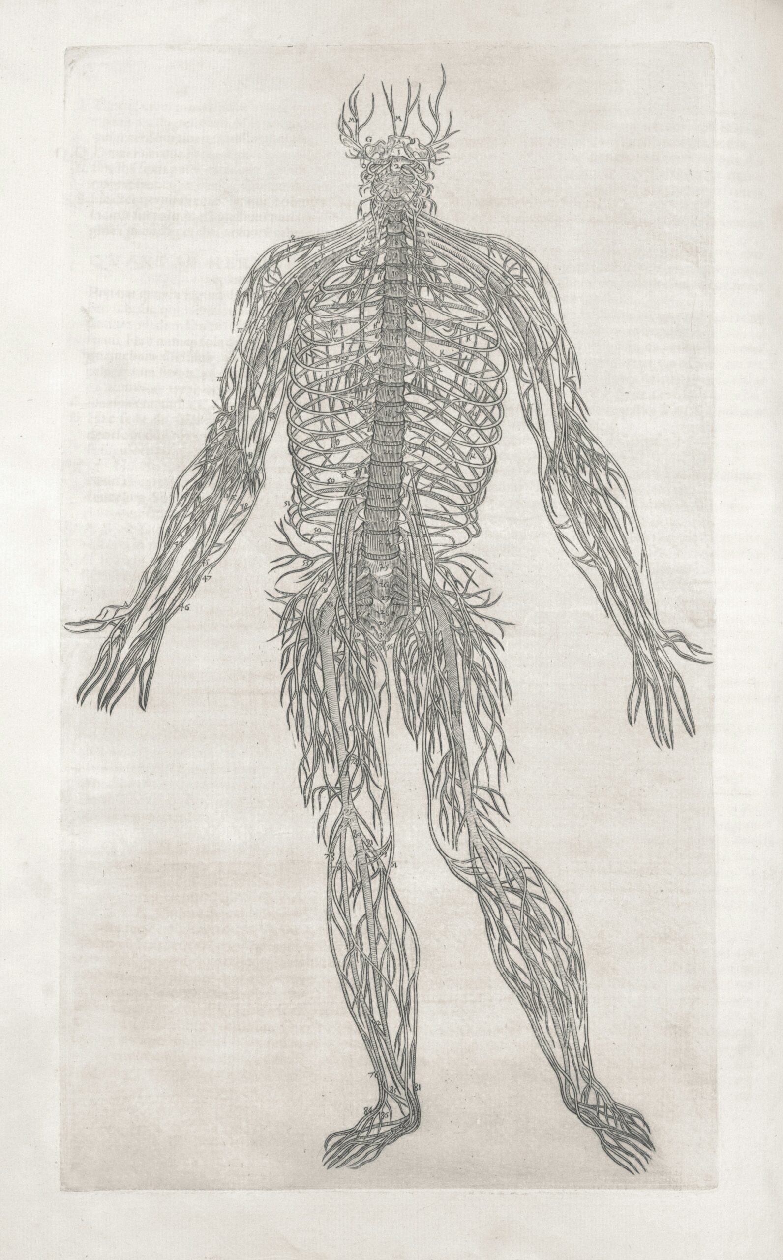
In order for the nervous system to utilize the information for mechanical force that is received from the senses, an interpretation of the received information must take place. This interpretation is made possible by proprioception, i.e. the information about position, movement, and amount of force required for completion of a movement that is continually received by the neuro-myo-fascio-skeletal system in order to enable said system to move in a coordinated fashion and to complete a wide variety of tasks.
The information gathered from these receptors can be distributed throughout the muscles, tendons, ligaments, and joints of the body. These sensory receptors allow the nervous system to gather information such as the length of the muscle, the rate of change of length of the muscle, the amount of tension in the tendons, and the amount of stress placed on the joints (movement and contact forces).
So long as a person has movement sense (proprioception) constantly organizing movement for them, then it seems easy enough to allow others to move.
Pathomechanics or abnormal movement patterns often seem to be a localized problem, i.e. a problem with one joint, one muscle or even part of a muscle. However, problems with a single joint or even a single muscle are often due to a number of factors. These include the distribution of force through tissues, the motor control of adjacent segments, the postures of adjacent joints and the mechanics of adjacent parts. In many cases a single factor can be identified as the cause of a problem; however, the problem can be distributed through a number of tissues that are in turn controlled by a number of different motor units, and therefore can affect a number of different joints.
This information about how the body does mechanotransduction can not be separated from information about how the nervous system does proprioception and reads movement.
Mechanotransduction: From Mechanical Input to Cellular Response
Mechanotransduction is where biomechanics and cell physiology meet. It describes how cells sense mechanical input and convert that input into biochemical activity. For movement professionals, the concept helps connect what can be seen from the outside, such as posture, joint motion, tissue tension, and load transfer, with what is happening on a much smaller scale inside responsive tissues.
Mechanical signals begin as physical events. A tendon is tensioned. Fascia is stretched or compressed. A joint surface receives pressure. Muscle fibers experience changes in length and load. Cells within these tissues are not separate from that environment. They are attached to neighboring cells and the extracellular matrix, and those attachments help carry mechanical information through the tissue.
In skeletal muscle, mechanotransduction refers to the process by which force and deformation are transmitted through muscle cells and their surrounding matrix, connecting the mechanics of movement with cellular-level response. Direction, intensity, repetition, rest, hydration, tissue state, and regional coordination all shape the mechanical environment that cells experience.
This is one reason kinesiology cannot be reduced to isolated joint actions. A shoulder abduction pattern involves the glenohumeral joint, scapulothoracic motion, clavicular movement, thoracic position, cervical tone, fascial continuity, and neural input. At the same time, tissues within that pattern are receiving mechanical information. The visible movement and the cellular environment belong to the same system.
Mechanotransduction gives pathomechanics another layer of meaning. Inefficient movement is more than poor alignment or weak muscles. It can also reflect how tissues are being loaded, unloaded, compressed, tensioned, and asked to coordinate over time. Understanding those forces helps practitioners think more clearly about movement quality, tissue behavior, and the relationships that shape normal function.
Cellular Signaling Language Outside the Classroom
Mechanotransduction gives movement professionals a practical way to connect mechanical loading with normal cellular activity. Terms such as cellular signaling, tissue responsiveness, matrix behavior, and biochemical communication show up in anatomy, exercise science, manual therapy, nutrition, and dietary-supplement discussions. The key is using each term in the right context.
In musculoskeletal education, cellular signaling is part of normal physiology. Cells respond to mechanical and chemical environments as part of ordinary biological activity, which is relevant to kinesiology because movement changes the mechanical environment of muscles, fascia, tendons, joints, and connective tissue.
Because these terms also appear outside formal anatomy education, movement professionals may encounter broader dietary-supplement discussions where consumers explore Khavinson peptide bioregulators alongside other supplement categories.
That distinction matters. The focus here remains anatomy, physiology, kinesiology, and pathomechanics. Supplement language should stay neutral and educational, without implying effects on muscle action, fascial mechanics, movement quality, tissue adaptation, or pathomechanical change.
Pathomechanics: When Load Distribution Becomes Less Efficient
When assessing a client it is often found that one joint, one muscle, etc. is not moving as it should in a certain range of motion. The causes for this decreased range of motion and resulting less than optimal movement pattern are distributed throughout the neuro-myo-fascio-skeletal system. Thus, when assessing a client for pathomechanics, one must consider the distribution of force during movement, the amount of motion that is being borrowed from adjacent regions of movement and the mechanical input to tissues during movement.
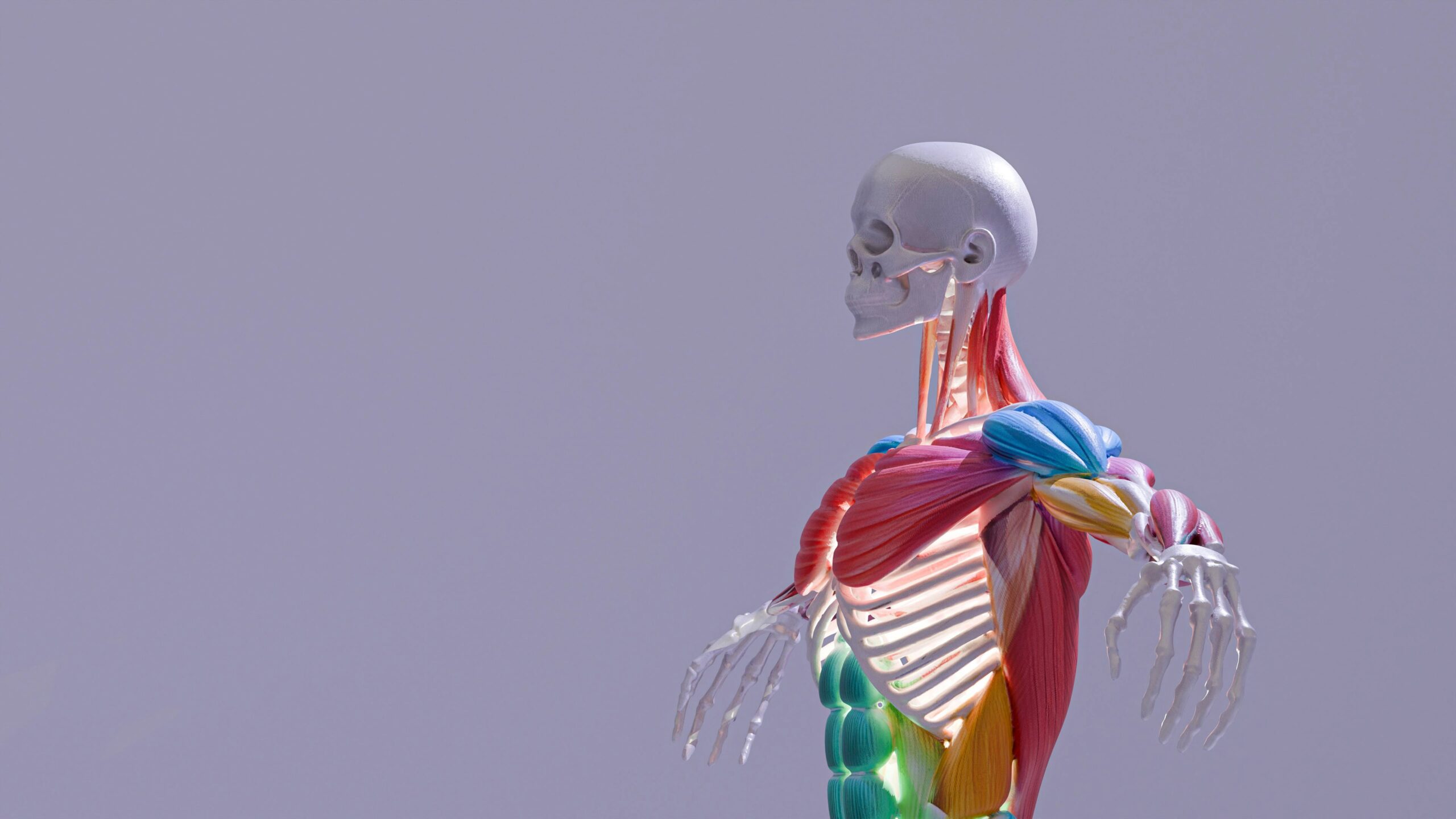
A decrease in joint movement can be caused by more than the joint itself. For example, the hip joint may have limited joint motion due to the position of the pelvis, restricted fascia, poor motor control and/or poor foot mechanics. The shoulder has limited glenohumeral joint movement but the scapula, thoracic spine and even the cervical spine are all involved. In many cases an overactive spinal segment appears to be overactive but in reality it is underactive due to compensatory patterns of movement in surrounding tissues.
Tissues experience repeated compression, tension, and shear as well as decreased movement variability when a person is in a posture or completes activities that expose them to loads. This mechanical environment of cells within various soft tissues affects how these cells sense their environment. Thus, for manual and movement professionals, understanding the structure of cells within various soft tissues and how these cells sense their environment is critical to their study of anatomy and physiology in relation to the biomechanics of the various tissues of the neuro-myo-fascio-skeletal system.
A simple assessment of tension in a muscle is not enough for the manual and movement therapists. They need to understand how force is distributed through the system. They also need to be able to recognize situations where a body is using excess motion and situations where a body is using less than necessary motion for a given task. Finally, they need to be able to recognize situations where tissues are being placed under increased mechanical loading than normal for a given movement.
Studying Pathomechanics of regions of the body that are not moving efficiently in a healthy manner is grounding in Anatomy and Physiology for the Manual and Movement Professionals. It enables them to study the mechanical forces placed on tissues of the body and how these tissues respond to these forces in a healthy manner. Thus they can gain insight into less than optimal movement patterns of healthy humans.
What This Means for Manual and Movement Professionals
A study of the Anatomy of the human muscles (with details of their origin, insertion and action via their appropriate innervation) of the human joints (with details of their appropriate axes, planes and arthrokinematics and available range of movement) of the human connective tissues (with details of the human fascia by continuity, glide, tension and regional inter relationships) is enhanced by viewing the human Anatomy as part of a dynamic communication system.
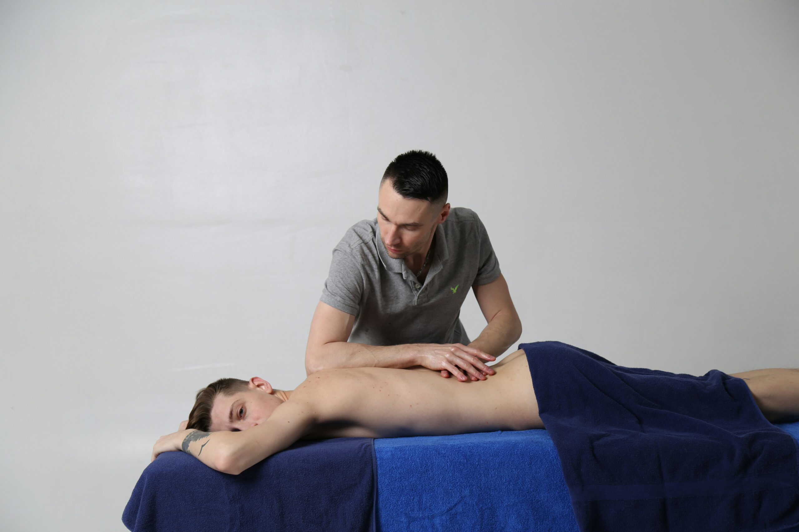
For manual therapists, trainers, Pilates and yoga instructors, it is very useful to look at a movement or at a person from the perspective of the neuro-myo-fascio-skeletal model. This will enable them to better assess a client and to better teach a person to move.
In terms of manual therapy of a local restriction, there are a number of layers to a movement. All of these need to be considered when assessing a movement or restriction and it is possible that a local restriction may have regional consequences. The person’s compensation for a restriction will be made up of a number of components including timing, proprioception, force distribution and tissue loading. The muscle that appears to be overactive may in fact be part of a larger movement strategy that is controlled by the whole of the neuro-myo-fascio-skeletal system.
In summary, there are many relationships that need to be further investigated. Forces are distributed throughout the entire muscle and the cells in the tissue experience these forces. Thus, a better understanding of the continuum of the neuro-myo-fascio-skeletal system will aid in our understanding of human movement from an anatomical, physiological, kinesiological and pathomechanical perspective.
Bringing the System Back Together
Mechanotransduction helps explain why movement education becomes more useful when anatomy and physiology are studied together. A joint action is never only a joint action. A muscle contraction is never isolated from fascia, tendon, sensory feedback, and the mechanical environment around the tissue. Each layer contributes to how the body organizes force.
This is where the neuro-myo-fascio-skeletal model becomes especially valuable. It gives practitioners a more complete language for what they already observe: muscles coordinating with fascia, nerves interpreting load, joints shaping motion, and tissues responding to the demands placed on them.
For students and professionals, the goal is not to make movement more complicated. The goal is to see the relationships clearly. Mechanotransduction offers a bridge between the mechanics of kinesiology and the physiology of living tissue, giving pathomechanics a broader and more connected frame.