
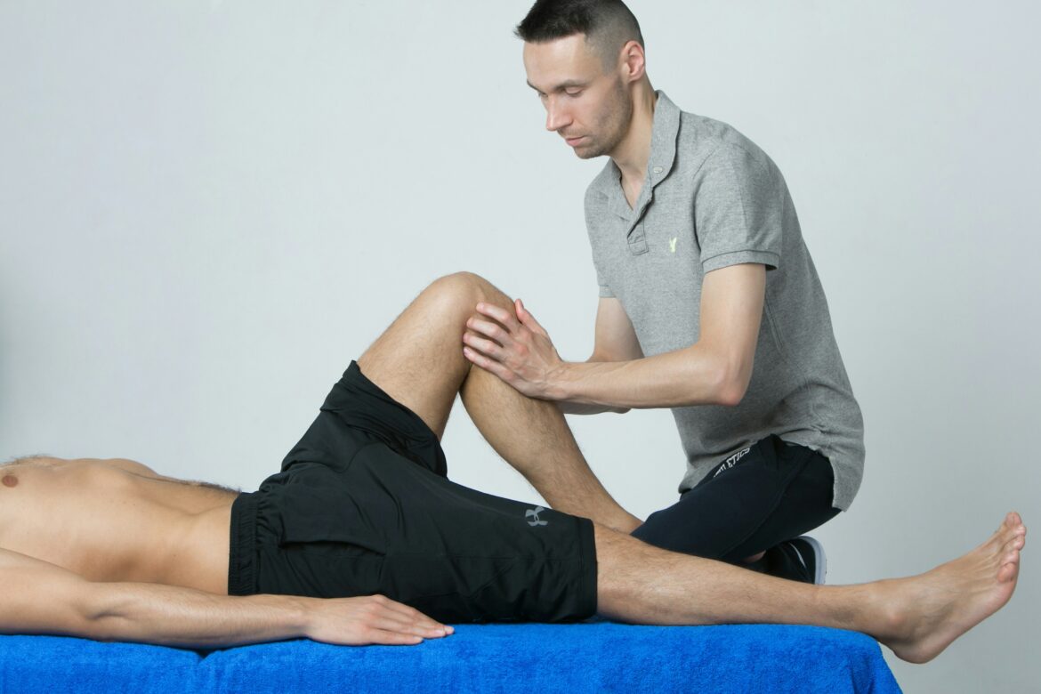
Reconstruction of the ACL is a common injury in sports that involves a lot of bending and twisting and the majority of these reconstructions are done arthroscopically. In general, most practitioners encounter a few clients with ACL’s that have been reconstructed and in reading the surgeons notes they can be quite bewildering. In general the surgery and subsequent rehabilitation is a long process during which time the tissues take a long time to come back to full strength. In manual therapy, finding surgical scars is common especially on the anterior aspect of the knee and often clients with reconstructed ACL’s have quite atrophied quadriceps.
It is very important to have a working knowledge of the ACL reconstruction surgery that has taken place and the subsequent changes of the soft tissues during the various healing phases when treating a client with an anteriorly located surgical scar on the knee or weakened and shrunken quadriceps muscles.
The Ligament and What It Actually Restrains
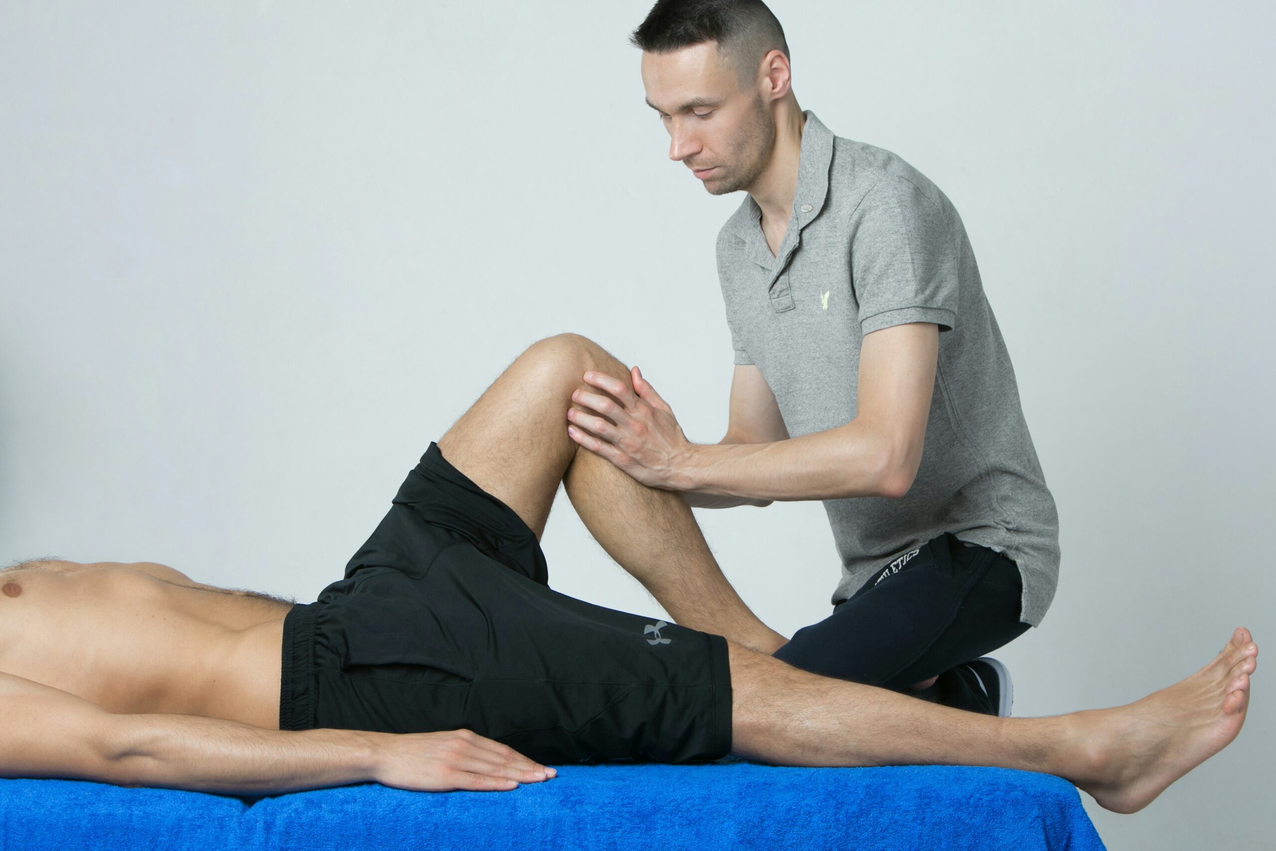
The ACL runs from the posterior aspect of the lateral femoral condyle to the anterior intercondylar area of the tibia. It is an intra-articular but extrasynovial ligament. The process of healing an ACL injury involves a lot of complex tissue changes. Understanding what actually happens in a joint with an ACL injury can be very useful when applying M.T. to a knee with such an injury.
The ACL restricts the anterior translation of the tibia in relation to the femur and, as well as this, it also allows the knee joint to deal with a large amount of rotational stress placed upon it. The primary function of the ACL, that of most importance to manual therapists, is that of preventing excessive internal tibial rotation. The patient with an ACL-deficient knee will frequently report a feeling of the knee ‘giving way’. This sensation is not necessarily painful and is seen to occur during activities that require a large amount of pivoting or change of direction. The patient may hear a popping sound at the time of injury, but this is not seen in all cases. As a result, the patient’s main complaint is not one of pain, but one of loss of function.
The ACL is a highly innervated structure. The large numbers of mechanoreceptors (sensory receptors) within the ligament provide information about the position of the knee. This information enables the surrounding muscles to make fine-tuned adjustments to protect the joint from injury. The client with an ACL deficient knee may report the knee ‘giving way’ during sport but report no pain. The giving way is due to a failure of the joint rotation (the knee has internally rotated) during movement such as landing from a jump. This is in no way related to pain.
How the Tear Typically Happens
The mechanism is more often non-contact than most people assume. As the National Institute of Arthritis and Musculoskeletal and Skin Diseases notes in its overview of sports injuries, ACL injuries are especially common in athletes and frequently occur when a person changes direction suddenly or lands from a jump.
The typical presentation involves deceleration with the knee near extension, a valgus moment, and internal rotation of the tibia beneath a relatively fixed foot. Clients often describe an audible pop, rapid effusion within the first few hours reflecting haemarthrosis, and an inability to continue the activity. Concurrent injury is common rather than exceptional, with meniscal tears and collateral ligament involvement frequently accompanying the ACL rupture, which is worth remembering when a client’s presentation seems more complicated than a single-structure injury would suggest.
Why the Ligament Does Not Simply Heal
This is the question clients ask most often, and the answer is anatomical. Tissues that live inside a joint, bathed in synovial fluid, generally fail to heal spontaneously after injury. The environment does not permit the organized clot formation that initiates repair in extra-articular tissue, and the torn ends retract and are eventually resorbed.
That biological reality is why reconstruction, rather than repair, has been the standard surgical approach. The ligament is not sewn back together. It is replaced.
What Reconstruction Actually Involves
The surgeon drills tunnels through the tibia and femur at the anatomical attachment sites and passes a graft through them, fixing it at both ends. Graft options fall into two broad categories. Autografts use the patient’s own tissue, most commonly bone-patellar tendon-bone, hamstring tendons, or quadriceps tendon. Allografts use donor tissue, avoiding donor-site morbidity at the cost of slower incorporation.
Graft selection is a genuine clinical decision rather than a matter of surgeon preference alone, weighing the patient’s age, sport, occupation and previous surgical history. Surgeons who handle high volumes of these cases, including revision procedures where a previous reconstruction has failed, tend to make that decision differently from those who perform them occasionally. A practice such as that of Steven Thornton ACL surgeon Dallas TX, a board-certified orthopedic surgeon with fellowship training in sports medicine at the Hospital for Special Surgery, illustrates the pattern: alongside primary reconstruction sit revision ACL work, meniscal root repair and cartilage restoration, procedures that cluster together because the knees requiring them frequently present with more than one problem.
For the manual therapist, the practical implication is that graft choice determines which donor site is also healing. A client with a patellar tendon autograft has an anterior knee that is sore for reasons unrelated to the reconstruction itself, and a hamstring graft leaves posterior thigh tissue that will influence what you find on palpation for months.
The Quadriceps Problem
If there is one phenomenon worth understanding above all others, it is arthrogenic muscle inhibition. Following knee injury and surgery, the quadriceps, and particularly vastus medialis, undergoes reflexive neural inhibition that is not simply disuse atrophy. The muscle is not merely weak from inactivity; the nervous system is actively limiting its recruitment in response to joint effusion and altered afferent input.
This matters clinically because it explains why strength returns slowly and unevenly even in motivated clients, and why extension deficits persist. It also means interventions that reduce effusion and normalize afferent input from the joint may facilitate recruitment in ways that loading alone does not. Persistent quadriceps deficits are among the better-documented predictors of poor functional outcome, so this is not a peripheral concern.
Where Hands-On Work Contributes
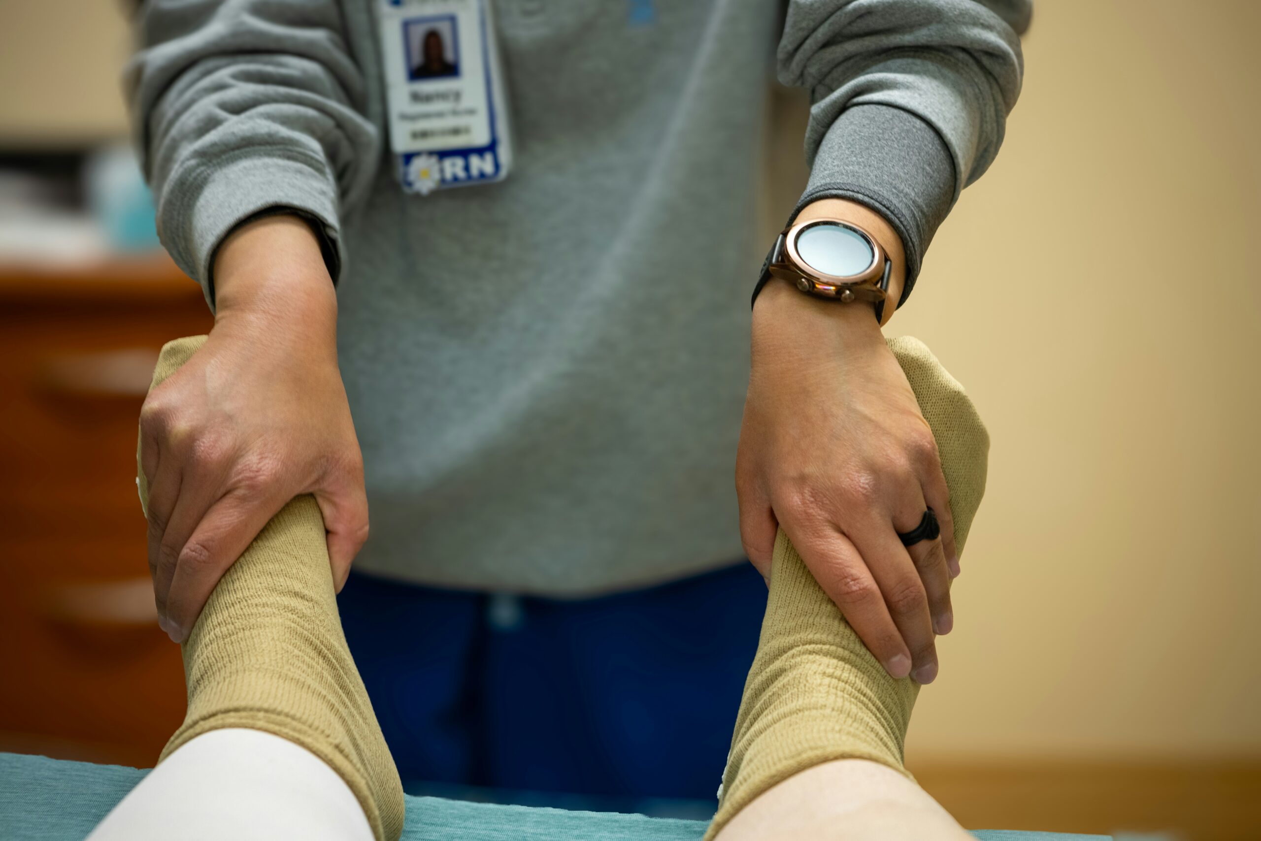
Several areas fall comfortably within manual therapy scope and genuinely influence outcome.
Scar mobility is the obvious one. Portal sites and incisions can tether superficial tissue to underlying structures, and restricted scar mobility around the anterior knee can limit patellar movement. Patellar mobilization deserves attention in its own right, since restricted superior glide will limit terminal extension and restricted inferior glide will limit flexion, both common in the months following surgery.
Beyond the knee, look up and down the chain. Hip abductor and external rotator function influences frontal-plane knee control, and clients who tore an ACL through a valgus mechanism frequently demonstrate hip weakness that preceded rather than followed the injury. Ankle dorsiflexion restriction alters landing mechanics and loads the knee differently. Neither is treated by working on the knee alone.
Compensatory patterns in the uninvolved limb also warrant attention, since months of asymmetric loading leave their mark on the other side, and clients rarely mention it.
The Timeline That Frustrates Everyone
The graft will go through a number of stages including necrosis, revascularization and remodeling. This process will take a number of months for full incorporation and the knee must be returned to full activities slowly and within the restrictions of the rehabilitation program.
When the knee is 3-6 months post operatively it is usually in the late phase of graft incorporation. The graft has started to take on the properties of a ligament and the knee is usually getting stronger. It is at this time that manual therapy can start to have the biggest impact on the knee. The practitioner needs to understand that the knee is still a long way off from full recovery and is still very prone to injury. It is at this time that many athletes start to return to sport and return to high levels of activity. They often believe that their knee is ready for this level of activity and are often shocked and disappointed when they are unable to perform at the expected level.
Working Alongside the Surgical Team
The manual therapist’s position is to work as a collaborating health care provider, within the client’s treatment plan, alongside the client’s orthopedic surgeon and physical therapist. This position requires the manual therapist to have knowledge of the type of graft used in the client’s ACL reconstruction (patellar tendon, semitendinosus and gracilis, quadriceps snip, etc.). Additionally, the manual therapist must have knowledge of the restrictions on the client’s knee following surgery, and how long it has been since surgery (3 months, 9 months, etc.). The restrictions on the knee and the length of time since surgery are much more important than the loading of the knee and return to sports in the manual therapist’s assessment of whether to treat the client’s knee. Working along side the orthopedic surgeon and physical therapist, within the client’s treatment plan, the manual therapist can do a lot of good for the client’s knee.
This article is provided as general professional information. It is not intended to be used for individual treatment of a client who has had an ACL reconstruction. Such clients must be treated within the constraints of their surgeon’s aftercare protocols. However, there is much that can be done with such patients, and those who have a good working knowledge of the surgery will find that they can treat these clients a lot faster than those who work only from the appearance of the patient’s scar.
Written by Lea Collins