
Signs and symptoms of overpronation (dropped arch / flat foot):
The first and most obvious sign of overpronation is a flat foot/dropped arch. The supple flat foot will have an arch when not weight bearing and will be seen to lose the arch upon weight bearing. A rigid flat foot will be flat whether the person is weight bearing or not weight bearing. Because overpronation results in medial rotation of the talus, leg, and thigh, the client’s/patient’s lower extremity will usually excessively medially rotate when standing.
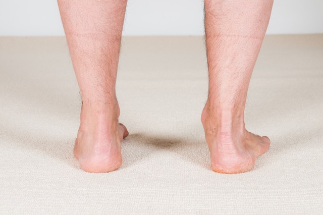
Posterior view of overpronation of the right foot. Permission: Joseph E. Muscolino.
Pain does not necessarily accompany this condition, but it often does. A supple flat foot results in the arch excessively dropping each time the foot strikes the ground. This causes the soft tissues on the underside of the foot to be forcefully stretched each time, tugging at their attachments and likely causing either spasms in the plantar intrinsic musculature (due to the muscle spindle reflex) and/or inflammation of the plantar fascia, known as plantar fasciitis. Either of these conditions can cause pain, especially upon weight bearing. Because these tissues attach to the underside of the calcaneus, the stretching forces placed upon them will be transmitted to the calcaneus, possibly eventually leading to a heel spur (due to Wolff’s Law: the deposition of calcium in response to physical stress). Therefore, overpronation is often accompanied by plantar intrinsic musculature spasm, plantar fasciitis, and heel spur.
Overpronation can also cause ramifications farther up the client’s/patient’s body. Dropping the arch tends to increase genu valgus posture, which places increased tension stress to the medial knee and increased compression stress to the lateral knee. Further, if the overpronation is present on one side only, or if it is present to a greater degree on one side than the other, then the pelvis on that side will drop. This places an asymmetrical force on the client’s/patient’s sacroiliac joints and also often results in a compensatory scoliosis to bring the head back to level.
Assessment/Diagnosis of overpronation (dropped arch / flat foot):
Assessment/diagnosis of overpronation follows from the signs and symptoms of this condition. The most important assessment tool is static or dynamic postural assessment, which will reveal the characteristic dropped arch. For static postural assessment, have the client/patient stand facing you, a few feet away, and note the height of the arches, including the relative symmetry from left to right side. Note also the orientation of the patella on each side. Patellar orientation will follow the rotation of the thigh; with overpronation, the patella on that side will be oriented more medially. If a dropped arch is found, postural examination should also look to correlate the presence of genu valgus.
Static postural assessment can also be done from the posterior perspective. In this case, instead of viewing the arches directly, look at the Achilles’ (calcaneal) tendons; each tendon should be vertical. With a collapsed arch, the Achilles’ tendon will bow inward instead. If the dropped arch is unilateral or greater on one side than the other, postural examination should include evaluation of a dropped iliac crest and possible compensatory scoliosis as well.
Dynamic postural assessment can be even more effective. With the client/patient facing you, ask the client/patient to march in place. It is important that the client/patient moves slowly and lifts each foot high enough (close to the height of the other knee) so that you have time to observe how much the weight-bearing arch drops on each side. If you have enough space, for example a long hallway, the client/patient can be asked to walk away and toward you, while you observe the arches, patellae, and Achilles’ tendons.
When evaluating pronation motion, keep in mind that upon weight bearing when standing, marching, or walking, the foot should pronate to some degree, and therefore the arch should drop somewhat. Because there is not universal consensus on exactly what subtalar joint neutral posture is, and exactly how much pronation is healthy versus unhealthy, it is best to eyeball this motion, using your best judgment. It is also helpful to compare left and right sides; symmetry should be present.
Palpatory examination should also be done to check for the presence of tightness and/or myofascial trigger points (TrPs) in the associated musculature. It is important to check all muscles that help to support the arch of the foot because they might develop TrPs as they attempt to control the excessive pronation. Similarly, palpate the antagonists to these muscles to see if their baseline tone is contributing directly to the overpronation. Palpatory examination should also be performed to assess for fascial adhesions within the plantar surface of the foot. Generally speaking, the more fascial adhesions, the more “rigid” the foot is. After palpatory examination of the soft tissues, it is important to assess for joint play/mobilization of the joints of the foot, especially assessing the motion of the bones/joints to move from plantar to dorsal in direction.
Range of motion of the foot at the subtalar and ankle joints should also be done, with particular attention to the client’s/patient’s inversion and eversion ranges; inversion is often limited in clients with overpronation. It is also important to conduct a verbal history to determine whether the client/patient has any habitual postures that might contribute to overpronation. For example, sitting cross-legged with the ankle of one leg placed on the thigh of the other, or driving with the heel of the right foot placed in front of the brake and the thigh turned out so that the toes of the foot are on the gas pedal. These postures tend to promote a turned out posture of the foot. Checking the client’s/patient’s shoes for excessive wear on the medial side of the heel can also be helpful.
Overpronation is a dysfunctional postural condition of the musculoskeletal system, so no further medical diagnosis/assessment is needed. However, if X-Rays are done, they can support the assessment by showing the dropped posture of the bones of the foot; these films should be done weight bearing, unless a rigid flat foot is being assessed, in which case non-weight bearing films could be done. X-Rays could also be done to determine the posture of the client’s/patient’s knee joints, pelvis, and spine, to determine what affect, if any, the overpronation has on the joints above.
Differential assessment of overpronation (dropped arch / flat foot):
When a client/patient presents with overpronation, it is important to differentially assess whether it is due to a rigid or supple flat foot. It is also important, as mentioned, to assess the possible presence of the postural affects of overpronation higher up in the body. Look especially for genu valgus, medially rotate thigh, dropped iliac crest on the side of overpronation, and a possible compensatory scoliosis.
