Signs and symptoms:
The most common symptom of iliotibial band friction syndrome (ITBFS) is pain in the distal lateral thigh over the lateral epicondyle of the femur. Swelling is also often present. The pain and swelling are usually exacerbated with activities that increase the rubbing of the ITB over the underlying bony landmark, such as running or cycling. When running, pain is experienced during the gait cycle on the stance limb side. When cycling, pain is experienced on the side that is pressing against the pedal.
Assessment/Diagnosis:

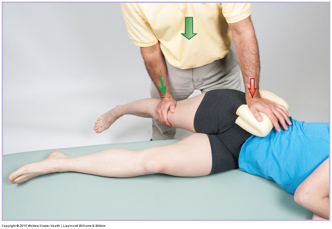
Ober’s test for iliotibial band friction syndrome. Permission:Joseph E. Muscolino. Manual Therapy for the Low Back and Pelvis: A Clinical Orthopedic Approach (2015).
Assessment/diagnosis of ITBFS is fairly straightforward. Pain and/or swelling can be palpated in the distal lateral thigh on the ITB at the location of the lateral epicondyle. Ober’s test can also be used to assess ITBFS. With the client/patient side-lying and oriented diagonally on the table, bring the client’s/patient’s thigh slightly into extension and then down into adduction at the hip joint, stretching the ITB and pulling it taut over the lateral femoral epicondyle. Pain at either of this bony landmark is positive for the condition. Noble compression test can also be used. Begin with the supine client’s/patient’s hip and knee joints passively flexed to 90 degrees. While palpating on the ITB at the lateral epicondyle of the femur, ask the client/patient to actively extend their hip and knee joints. This test is considered positive if the client/patient experiences pain under your palpating fingers at approximately 20-30 degrees of knee joint flexion. Given their role in ITBFS, it is also important to palpate the client’s/patient’s tensor fasciae latae, gluteus maximus, and vastus lateralis muscles.
Because ITBFS is often caused or aggravated by the presence of other musculoskeletal conditions, it is important to also assess for these conditions. They include genu varum, genu valgum, and overpronation of the foot; these conditions can be evaluated with postural exam. As a part of genu valgum assessment, check for the strength of the gluteus medius by having the client/patient stand on one limb; if the client’s/patient’s gluteus medius is weak, their pelvis will drop to the opposite side. When assessing overpronation of the foot, check for the leg (tibia) and thigh (femur) to fall into medial rotation when weight bearing. This can usually be best seen by observing the posture of the patella.
Differential assessment:
Local pain directly over the lateral femoral condyle is a fairly clear indicator of ITBFS. However, differential assessment should be done for a few other conditions. If pain is located near the lateral femoral condyle, but more distally over the knee joint, it is important to differentially assess for conditions of the knee joint such as a tear to the lateral meniscus, a sprain of the lateral collateral ligament, or degenerative joint disease (osteoarthritis). Pain in the lateral thigh over the ITB between the lateral condyle and greater trochanter is often due to tightness in the underlying vastus lateralis or perhaps vastus intermedius musculature and does not necessarily indicate pathology of the ITB. Pain located directly over the greater trochanter is often indicative of greater trochanteric bursitis. And pain proximal to the greater trochanter is likely due to myofascial trigger points in the gluteus medius.
