Introduction
Leon Chaitow recently wrote an editorial in the Journal of Bodywork and Movement Therapies asking which would be the better term to describe the gentle soft tissue manipulation approach usually referred to as myofascial release: Myofascial Release or Myofascial Induction.
Myofascial Release and Myofascial Induction
The technique of Myofascial Release (MFR) has its roots in osteopathy. Carol Manheim wrote that although many osteopaths had used and taught these soft tissue stretching techniques, it was not named until the 1980s when Dr. Robert Ward taught it as an independent course and used the term Myofascial Release.
Meanwhile, Myofascial Induction Therapy (MIT) is a relatively new term introduced by Andrzej Pilat, a physiotherapist (physical therapist) from Madrid, Spain. While the two approaches are basically the same, Andrzej stressed that “induction” is related to the facilitation of movement rather than passive stretching (or release) of the fascial system.
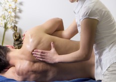
Techniques used in either MFR or MIT usually involve:
- Therapist places hand(s) on tissues to be treated
- Presses lightly and engages ease or bind barriers
- Applies a constant pressure to tension the tissue
- Patient participation may enhance the process
- After overcoming each barrier, pressure application can be modified depending on tissue resistance
- Pressure should be reduced if there is any pain
Research Study
In a systematic review of 19 randomized, controlled trials, involving MFR (out of 133 studies evaluated), Ajimsha et al. (2015) found that “MFR is emerging as a strategy with a solid evidence base and tremendous potential.“ MFR was defined in this systematic review as:
“…a form of manual therapy that involves the application of a low load, long duration stretch to the myofascial complex, intended to restore optimal length, decrease pain, and improve function.“
Andrzej Pilat wrote:
“The term ‘induction’ relates to the correction of movement facilitation, and not a passive stretching of the fascial system. This is primarily an educational process, in the search for restored optimal homeostatic levels, recovering range of motion, appropriate tension, strength, and coordination. The final aim of the therapeutic process is not establishment of stable hierarchies, but facilitation of optimal and continuous adaptation to environmental demands, with maximum efficiency.”
He further explains the difference between induction, and release:
“Clinicians familiar with myofascial release (MFR) note the many similarities between it and MIT. With different nuances, they are based on the same concept of clinical reasoning and complement each other. MIT is characterized as manual tissue remodelling, always avoiding arbitrary stimulus application (altered force intensity and direction), focusing on the intrinsic natural tissue response.”

Mechanisms of Action
There are several hypotheses on the mechanisms by which MFR/MIT work:
Cellular Level
Some studies looked at the cellular level, by modelling the effect of MFR in vivo on fibroblast morphology and physiology. The basis is that repetitive motion strain is caused by cyclic short-duration stretches. The key components of the response to mechanical forces are fibroblasts, which are the main fascial cells that respond to different types of strain by secreting anti-inflammatory chemicals and growth factors, thus improving wound healing and myofascial repair processes.
For this particular purpose, myofascial release was described as an acyclic long-duration stretch (ALDS) to myofascia tissue to repair injury. Research on cellular application of “stretch” shows that fibroblasts respond to different types of strain by secreting anti-inflammatory chemicals and growth factors, thus improving would healing and muscle repair process. It also appears that the magnitude, direction, frequency, and duration of ‘strain’ affect fibroblastic response to would healing.
It is important to note that these studies are simulated in the laboratory in a petri dish; thus it is still uncertain how this would translate to real-life MFR application.
Neurophysiological Response
In addition to mechanoreceptor effects, proprioceptors, nociceptors, and interoceptors are likely to be stimulated during application of MFR/MIT.
Enhanced Fascial Gliding
At the muscular level, MFR is commonly believed to ease fascial gliding motion, releasing adhesions between fascia.
A study by Chen et al. (2016) demonstrated that in patients with chronic low back pain, light, 60-second sustained compression of the lumbar interfascial triangle (a facial intersection of fascia of the paraspinal musculature with thoracolumbar fascia, between the 12th rib and the iliac crest), produced demonstrable (via ultrasonography) improvement in the behavior the sliding function of transversus abdominis during contraction. They concluded that the study ”indicated immediate effect of sustained manual pressure on the musculofascial junction of transversus abdominis (TrA) – and that this supported the concept that the possible imbalanced tension of the myofascial corset of transversus abdominis in patients with LBP.”
Which Term is Better?
Based on the evidence and arguments presented, Leon Chaitow in his editorial concluded that myofascial induction would seem to be the more appropriate term.
This blog post article was created in collaboration with TerraRosa.com.au.
