
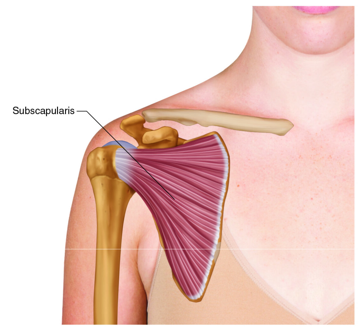
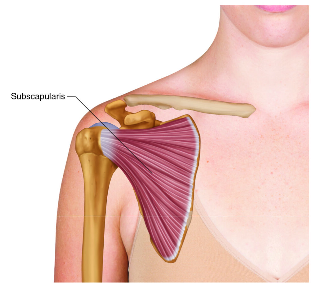
Subscapularis – one of the four rotator cuff muscles. Credit: Dr. Joe Muscolino (learnmuscles.com).
Introduction
The Subscapularis is one of the four rotator cuff muscles.
The name “subscapularis” tells us that it is located on the anterior side of the scapula (“sub” as in “under” from the typical perspective of viewing the scapula from the posterior side; hence it is on the anterior side).
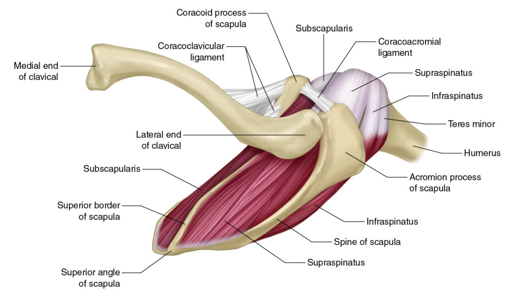
Superior view of the right scapula with the rotator cuff muscles. Credit: Dr. Joe Muscolino (learnmuscles.com).
The other three rotator cuff muscles are the supraspinatus, infraspinatus, and teres minor, which are all located on the posterior/dorsal side of the scapula.
Subscapularis Attachments
The subscapularis attaches from the:
anterior side of the scapula
to the
lesser tubercle of the humerus
Subscapularis Functions
The subscapularis is a medial rotator of the arm at the shoulder/glenohumeral (GH) joint. This is its concentric/shortening joint action function.
However, when the subscapularis is isometrically contracting, as with the rotator group as a whole, it functions to stabilize the head of the humerus into the glenoid fossa of the scapula.
This stabilization function is quite fascinating… Whenever the arm is lifted upward at the GH joint (whether it is flexion, extension, abduction, or adduction), the distal end needs to lift, but the proximal end, the head, needs to be stabilized down into the glenoid fossa. This isometric-contraction stabilization function is actually more important functionally than its medial rotation joint action function (given that there are other larger muscles like the latissimus dorsi, teres major, and pectoralis major that can all medially rotate the arm at the GH joint).
Nearby Anatomy
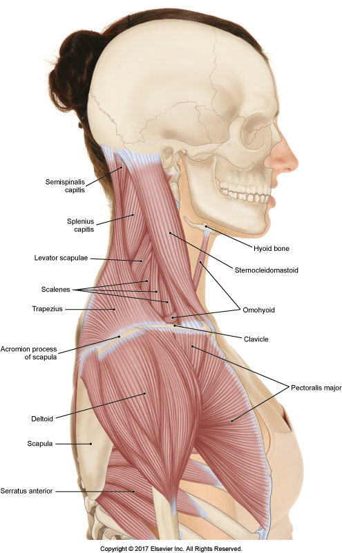
Right lateral view of the upper body. Credit: Dr. Joe Muscolino – The Muscular System Manual, 5th ed.
The subscapularis is in a fairly unique location, sitting between the scapula and the thoracic rib cage wall. One other muscle is also located in this space; that is the serratus anterior. The difference is that the subscapularis hugs the scapula, whereas the serratus anterior hugs the rib cage… so if you can physically open up the space between the scapula and rib cage wall, the subscapularis will be located posteriorly and laterally against the scapula and the serratus anterior will be located anteriorly and medially against the rib cage.
The distal attachment of the subscapularis is onto the lesser tubercle of the humerus, directly deep to the anterior deltoid.
All rotator cuff muscles attach distally onto the two tubercles of the humerus, so the other three rotator cuff muscles attach onto the greater tubercle.
Palpating the Subscapularis

Positioning to palpate the right-side subscapularis. Credit: Dr. Joe Muscolino – The Muscle and Bone Palpation Manual, 3ed.
The subscapularis can be a challenging muscle to palpate because it is so deep-seated, located between the scapula and thoracic rib cage wall. Palpating its belly requires the tissues of the client’s body in this region to be sufficiently mobile to allow the scapula to be separated from the thoracic rib cage wall. Given the challenge of accessing this muscle, it is extremely helpful to be able to visualize the location of the subscapularis.Visualization helps to guide your hands during the palpation protocol.
Palpation of the right-side subscapularis:
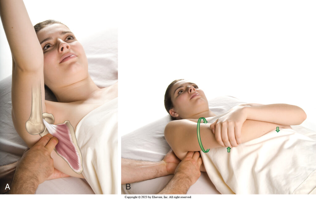
Visualization of (Figure A) and Palpation of (Figure B) of the subscapularis. Credit: Dr. Joe Muscolino – The Muscle and Bone Palpation Manual, 3ed.
Have the client supine with their right forearm resting across their abdomen. Reach in between the scapula and their thoracic rib cage body wall with your finger pads (leading somewhat with your fingertips) oriented laterally away from their body wall, aiming toward the anterior surface of their right scapula. Their left hand can help support the position of the right upper extremity.
The reason to have their right forearm resting on their abdomen is that it requires the arm to be adducted, which slackens the tissue in the area, thereby allowing better access deeper into the tissues to reach the subscapularis. However, the problem with this upper extremity position is that it obscures your ability to see where your palpating fingers are located and the direction in which they are sinking. This is why it is so important to be able to visualize the muscle.
To confirm that you have reached the subscapularis, ask the client to gently medially rotate their right arm at the GH joint by lifting their right elbow up away from their trunk, while keeping their right hand down on their abdomen. If you are on the subscapularis, you will feel it engage/pop.
Because the subscapularis is so deep, and because the palpation protocol somewhat requires leading with the fingertips (which can be pokier than finger pad palpations), and because it is so seldomly palpated, it is usually very sensitive to palpation, so it is extremely important to sink in very slowly, allowing the client a chance to accept the pressure. It is also important to work with the client’s breath… ask the client to breathe in, and then as the client breathes out, you breathe out with them (“sympathetic breathing”) and SLOWLY sink in. It might be necessary to perform three to four cycles of breath with a slightly greater depth of excursion into the tissues each time, until you have reached the muscle.
Be aware that there are occasional clients whose tissues between the scapula and rib cage are so adhesed that it is not possible to reach in with sufficient depth to reach the subscapularis. If this is the case, then it might be possible in time to access the subscapularis if the tissues in the region are gradually warmed up and made flexible over multiple sessions.
Massaging the Subscapularis
Massaging the subscapularis follows from the palpation protocol. Given the preparation and set up necessary to palpate this muscle, it is efficient to work it once it is located (assuming that it would benefit from the massage). Work SLOWLY and deliberatively, with successive excursions attempting to reach in farther toward the fibers closer to the medial border of the scapula.
To palpate and massage the distal tendon on the lesser tubercle of the humerus, keep working successively from the scapula in the medial direction toward the humerus, moving in baby steps with each successive excursion. It is usually necessary to change the position of their right arm to be up and supported by (resting on) your left shoulder so that you can access the humerus (however, keep in mind that this position might tauten the fascial tissues in the region, making it harder to sink in to the subscapularis distal humeral tendon).
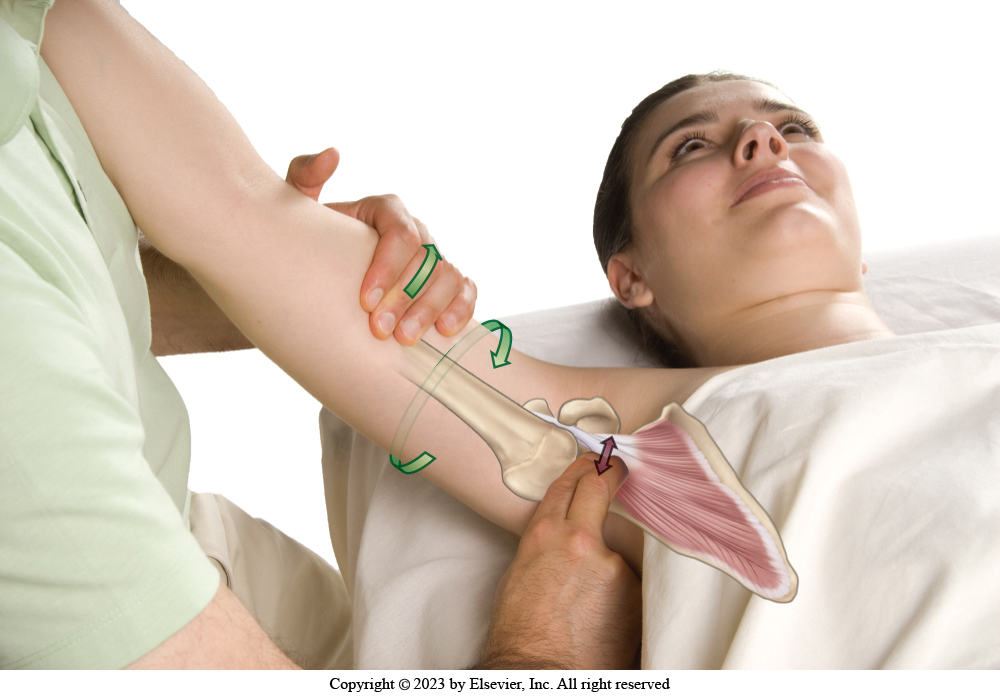
Palpating and massaging the humeral attachment of the subscapularis. Credit: Dr. Joe Muscolino – The Muscle and Bone Palpation Manual, 3ed.
If it is not possible to actually reach the attachment on the humerus with this protocol, then it is usually possible to work the distal tendon through the anterior deltoid. Do this by strumming vertically/perpendicularly across it.
Stretching the Subscapularis
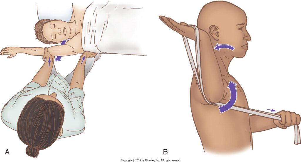
Stretching the right-side subscapularis. Therapist-assisted and client self-care. Credit: Dr. Joe Muscolino – The Muscle and Bone Palpation Manual, 3ed.
The subscapularis is a medial rotator of the arm/humerus at the GH joint, so it is stretched with lateral rotation. This is usually best accomplished with the help of a rope or band as seen in the accompanying illustration. If you do not have a rope or band, then using a doorframe might work… place your distal forearm against the doorframe to stabilize your arm, and turn/rotate your body in the opposite direction.
Subscapularis and Frozen Shoulder
The subscapularis is often implicated in frozen shoulder. Frozen shoulder is a condition that is marked by loss of range of motion of the arm at the shoulder joint. It often begins as a condition in which musculature tightens up, thereby limiting GH joint range of motion (this stage is often described as neurogenic frozen shoulder because it is caused by the nervous system ordering muscle contraction). If left untreated, if often becomes a condition marked by fascial adhesions in the ligament/joint capsule complex; this phase often described as adhesive capsulitis.
Frozen shoulder can be marked by loss of any GH joint range of motion, but most often it is characterized by a loss of abduction; and/or secondarily by a loss of lateral rotation. If the client has decreased lateral rotation, then medial rotation musculature, especially the subscapularis, will usually be tight and would benefit from manual therapy treatment, both massage and stretching.
Especially when frozen shoulder has reached the stage of adhesive capsulitis, it is often in need of joint mobilization, in which the scapula is stabilized and the humerus is mobilized relative to it. For manual therapy practitioners who are schooled in this technique, it is extremely valuable for the client with frozen shoulder because regular stretching alone usually cannot isolate the stretch force to the ligament/joint capsular tissues between the humerus and scapula because when the humerus is moved, the scapula moves with it. Hence the need for the scapular stabilization aspect of joint mobilization. Note: the type of joint mobilization usually employed here is Grade IV slow-oscillation joint mobilization (not Grade V fast-thrust).
Conclusion
The subscapularis is one of the more challenging muscles for manual and movement therapists to work with because it is so deep-seated, but given its role in frozen shoulder, it can be one of the more important muscles when working with our clients.
Biography

Dr. Joe Muscolino (learnmuscles.com).
Dr. Joseph Muscolino, DC is a soft-tissue oriented chiropractic physician and leading educator in manual and movement therapy. He is the author of eight major textbooks published by Elsevier and LWW, translated into more than 10 languages and used worldwide in core curriculum and clinical practice. A global lecturer and NCBTMB-approved CE provider, he offers COMT (Clinical Orthopedic Manual Therapy) certification workshops across the US and internationally. Visit his website at: LearnMuscles.com.
LearnMuscles Continuing Education (LMCE) is one of his online subscription platforms with over 4,000 video lessons for manual and movement therapy professionals, and more than 320 free NCBTMB-CE hours.
What is the subscapularis muscle? The subscapularis is one of the four rotator cuff muscles, located on the anterior (front) surface of the scapula, between the scapula and the rib cage.
What does the subscapularis do? It medially rotates the arm at the shoulder (glenohumeral) joint, and — just as importantly — it isometrically stabilizes the head of the humerus into the glenoid fossa during all arm-lifting movements.
Where does the subscapularis attach? It attaches from the anterior surface of the scapula to the lesser tubercle of the humerus.
Why is the subscapularis hard to palpate? Because it sits deep between the scapula and the thoracic rib cage wall, palpating it requires separating the scapula from the rib cage and confirming placement by feeling the muscle engage during gentle medial rotation of the arm.
How is the subscapularis connected to frozen shoulder? The subscapularis is a medial rotator and is commonly tight in frozen shoulder (adhesive capsulitis), especially when a client presents with restricted lateral rotation, making it a key muscle for massage, stretching, and joint mobilization treatment.
How do you stretch the subscapularis? Since it medially rotates the arm, it’s stretched through lateral rotation — typically using a rope or band, or by bracing the forearm against a doorframe and rotating the body away from it.