This blog post series of articles presented and went into some depth discussing six unusual suspect muscles, each one of which may be the underlying cause of our client’s condition. However, there are so many more unusual suspect muscles that could have been presented. Following is a brief discussion of a few more unusual suspects.
Semispinalis Capitis
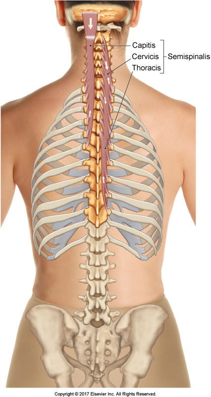
The semispinalis capitis of the posterior cervical spine is often overlooked as the cause of pain and tightness in the neck (Figure 20). It is actually the thickest muscle in the back of the neck and often tight and symptomatic. It lies directly deep to the upper trapezius so the upper trapezius is often blamed when the semispinalis is the offending culprit. When assessing clients with neck tightness and pain, look for the semispinalis. Deep work, over the laminar groove, directly lateral to the spinous processes, is often the key to helping clients with a tight semispinalis capitis.

Figure 20. Posterior view of the semispinalis group. Courtesy Joseph E. Muscolino. The Muscular System Manual – The Skeletal Muscles of the Human Body, 4ed. Elsevier. 2017.
Pectineus
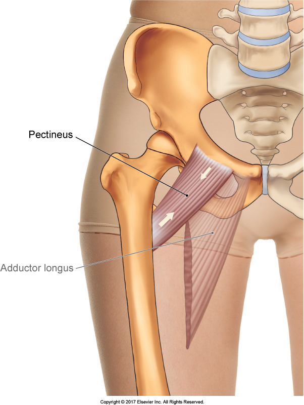
The pectineus is a transitional muscle between the hip flexor and hip adductor compartments; it is located between the psoas major of the flexor compartment and adductor longus of the adductor compartment (Figure 21). It is often missed because the therapist’s focus is so often on the psoas major. And because the pectineus sits a bit deeper than its neighbors, it is more challenging to find. The easiest way to locate the pectineus is to first find the proximal tendon of the adductor longus, and then drop immediately lateral to it. Keep pressure close to the pubic bone and ask the client to try to move the thigh at the hip joint against your resistance in an oblique plane that is a combination of flexion and adduction.

Figure 21. Anterior view of the right pectineus. Courtesy Joseph E. Muscolino. The Muscular System Manual – The Skeletal Muscles of the Human Body, 4ed. Elsevier. 2017.
Vastus Lateralis
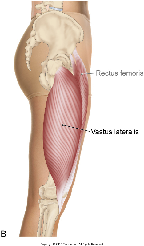
The vastus lateralis is a member of the quadriceps femoris group. As such, it is often thought of as being located only in the anterior thigh. However, the vastus lateralis attaches to the lateral lip of the linea aspera of the femur located all the way on the posterior side of the bone (Figure 22). Therefore, even though the vastus lateralis is superficial anterolaterally, it is also located in the lateral and posterolateral thigh. It is the lateral thigh where it is often overlooked as the causative agent of the client’s pain. Because the iliotibial band is located superficial to the vastus lateralis, the iliotibial band is often blamed as the cause of the pain when the deeper vastus lateralis is the true culprit. When the iliotibial band is the cause of the client’s condition, the pain will usually be felt at the lateral femoral condyle. If the client’s pain is anywhere mid-thigh, look instead to locate and assess the vastus lateralis. This is easy. To locate it, simply resist the client from trying to extend the leg at the knee joint and feel for the vastus lateralis to engage. Once located, have the client relax the muscle and assess its tone.
Note: An imbalance of strength between the vastus lateralis and the vastus medialis is a precipitating factor that can lead to patellofemoral syndrome.

Figure 22. Right vastus lateralis (lateral view). Courtesy Joseph E. Muscolino. The Muscular System Manual – The Skeletal Muscles of the Human Body, 4ed. Elsevier. 2017.
Note: This blog post article is the eighth in a series of 8 posts on
Unusual Suspect Muscles of the Body*
The 8 Blog Posts in this Series are:
- Introduction
- Palmar Interossei (PI)
- Flexor Pollicis Longus (FPL)
- Quadratus Femoris (QF)
- Coccygeus & Levator Ani
- Sternohyoid
- Longus Colli & Longus Capitis
- Other Unusual Suspects…
* This series of blog post articles is modified from an article originally published in massage and bodywork (m&b) magazine: The Unusual Suspects. November/December 2016 issue.


