Five Major Muscles of the Sacroiliac Joint Stabilization
This is the 1st of 3 blog post articles on the Muscles of Sacroiliac Stabilization.
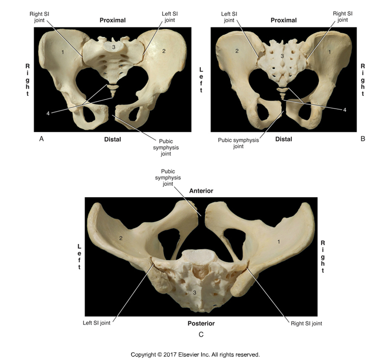
Sacroiliac Joint. Permission Joseph E. Muscolino. Kinesiology – The Skeletal System and Muscle Function, 3rd ed. (Elsevier, 2017).
Following are the five major muscle / muscle groups involved in sacroiliac stabilization that should be assessed and likely worked with manual therapy when the client presents with a sacroiliac joint condition.
- Piriformis
- Gluteus Maximus (superior deep fibers)
- Coccygeus and Levator Ani
- Paraspinals
- Hamstrings
Joints and Muscle Stabilization
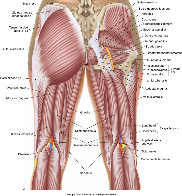
Musculature of the Pelvis and Hip Joint. Permission Joseph E. Muscolino. The Muscular System Manual – The Skeletal Muscles of the Human Body, 4th ed. (Elsevier, 2017).
When a joint has pain and/or dysfunction, it is common for the surrounding musculature to tighten in an effort to stabilize and protect the joint. This is certainly true when there is a problem with the sacroiliac joint (SIJ). So learning about the muscles of sacroiliac stabilization is valuable.
Sacroiliac Joint (SIJ) Conditions
If the SIJ is irritated and/or inflamed, in other words there is a ligamentous sprain, muscles tighten for sacroiliac stabilization, attempting to prevent the joint from excessive movement that might further irritate/damage it or cause pain to the intrinsic fascial tissue of the joint. If there is a muscular strain of the associated musculature of the SIJ, the muscles might be directly injured causing them to tighten up and armor the region. If instead, there is movement dysfunction, for example the SIJ is hypermobile, musculature might tighten to stabilize the joint to decrease its excessive movement. And even if it is hypomobile dysfunction, a hypomobile SIJ often does not move/glide smoothly and pain can occur during motion. Finally, if there is degenerative joint disease (osteoarthritis) of the SIJ joint surfaces, then musculature might tighten to minimize this motion and guard against the pain.
Chicken and Egg
And when the musculature remains too tight for too long, this often plays back on the joint, resulting in further pathomechanics. Tight musculature can excessively restrict the joint’s motion resulting in hypomobile dysfunction (which often causes the contralateral SIJ to become hypermobile as a compensation); after all, too much stabilization results in rigidity. And excessive muscular tightness can restrict free and easy gliding motion of the joint surfaces, resulting in irritation to the joint surfaces themselves. Further, tight musculature can cause pain in itself due to the excessive tension pulling forces of contraction upon its own fascial tissue. So we have a bit of a chicken and the egg scenario wherein the joint problem can lead to an external myofascial/muscular problem, which can then further the joint problem. Or the muscular problem can lead to the joint problem, which can then further the myofascial/muscular problem… chicken and the egg.
Reason to Treat
Regardless, of the underlying pathomechanics of the SIJ as the reason for the increased muscle tone, there is good reason for the manual therapist to know the muscles of sacroiliac stabilization that often respond to SIJ pain, injury, and dysfunction and become excessively tight. Even when the original reason for the sacroiliac stabilization and tightness is valid, often the human body overdoes it and tightens the musculature excessively and/or keeps it tight long after it needs to be, so the musculature becomes stuck in a chronic pattern of hypertonicity. So knowing which muscles to assess and how to work them if appropriate is valuable for the clinical orthopedic manual therapist.
Five Major Muscles /Muscle Groups of Sacroiliac Stabilization
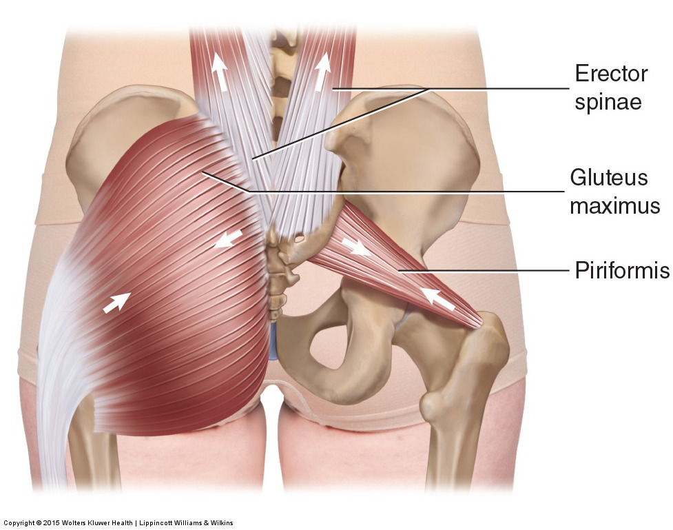
Musculature around the Sacroiliac Joint. Permission Joseph E. Muscolino. Manual Therapy for the Low Back and Pelvis – A Clinical Orthopedic Approach (2013).
Following are the five major muscles / muscle groups of sacroiliac stabilization that should be assessed and likely worked with manual therapy when the client presents with a sacroiliac joint condition.
- Piriformis
- Gluteus Maximus (superior deep fibers)
- Coccygeus and Levator Ani
- Paraspinals
- Hamstrings
This blog post article covers the Piriformis.
Piriformis
The piriformis attaches from the anterior sacrum to the greater trochanter of the femur. Therefore, it crosses the SIJ (as well as the hip joint) and is often one of the first muscles that will tighten when a SIJ injury/condition occurs.
Piriformis Palpation
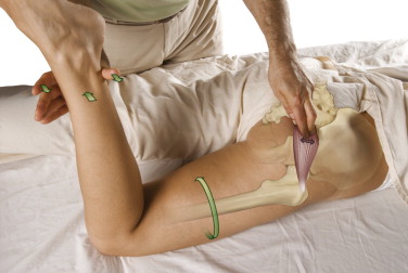
Piriformis Palpation. Permission Joseph E. Muscolino. The Muscle and Bone Palpation Manual – with Trigger Points, Referral Patterns, and Stretching, 2nd ed. (Elsevier, 2016).
To palpate and assess the piriformis, find the PSIS and the sacrococcygeal joint line, go halfway between them, and then drop laterally off the sacrum and you will be on the piriformis (deep to the gluteus maximus). Then draw a line from there to the superior aspect of the greater trochanter and that is the line of the piriformis. For confirmation, ask the client to laterally rotate the thigh at the hip joint (this is done with the client’s knee joint flexed to 90 degrees and resistance added to the medial surface of the distal [lower] leg) and feel for the contraction of the muscle. Once the location of the palpation is determined, palpate and assess it while at baseline tone.
This is the 1st of 3 blog post articles on Five Muscles of Sacroiliac Stabilization To Work On With Our Clients.
The 3 articles are:
(Click here for the blog post article: Piriformis Syndrome.)
