

Manual therapists spend a lot of time on the anterior neck. The scalenes and sternocleidomastoid come up in nearly every case of chronic neck pain, tension headache, and thoracic outlet symptoms. Most of us treat them well. Some clients still relapse within days.
One reason sits a few inches north of the tissue under your hands. A small group of muscles controls the width of the nasal airway. When those muscles cannot hold the airway open, the client shifts toward mouth breathing. That shift changes how the client breathes, and the anterior neck absorbs the extra work.
Surgeons who work on the nasal airway see the far end of this pattern. The Rhinoplasty Center of Long Island performs functional and cosmetic nasal surgery at an accredited surgery center in New York, and the practice says the complaint almost never arrives labeled correctly.
“Patients rarely walk in and tell us their nasal valve is collapsing,” the center says. “They tell us they sleep badly, they wake up with a dry mouth, and they cannot get a full breath on a run. Many of them have already spent a year with a therapist working on their neck.”
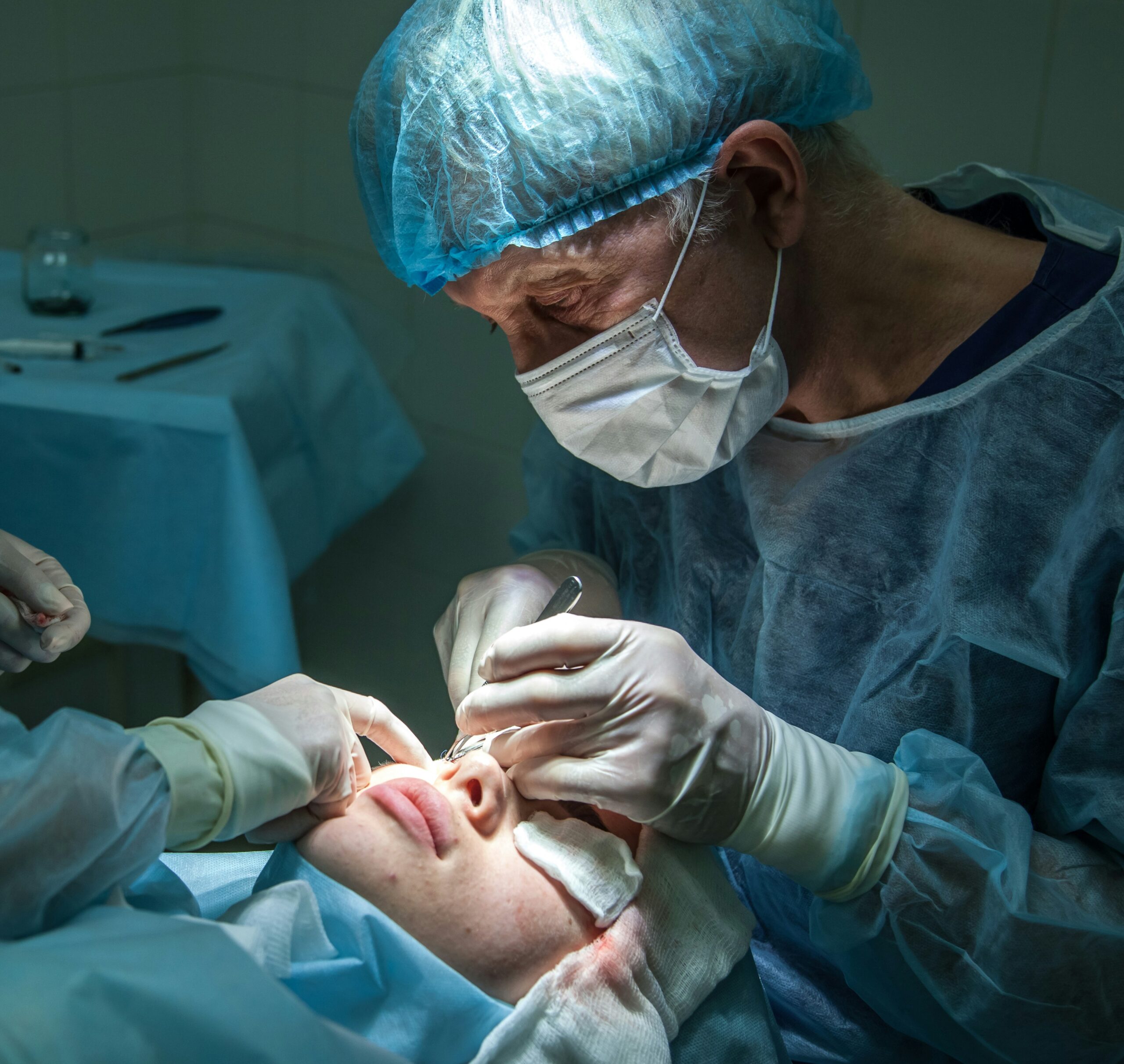
The nasal muscle group
These muscles rarely get more than a line in a palpation course. All of them derive from the second pharyngeal arch and receive innervation from the facial nerve.
Nasalis, transverse part: Also called compressor naris, this part runs from the maxilla above the canine fossa into a thin aponeurosis across the nasal dorsum. It compresses the nasal aperture and narrows the nostril.
Nasalis, alar part: Also called dilator naris posterior, this part attaches from the maxilla to the alar cartilage and the skin of the ala. It draws the ala laterally and widens the nostril.
Dilator naris anterior: This muscle arises from the lateral crus of the major alar cartilage and the adjacent accessory cartilage, then attaches into the skin at the alar groove. It opens the nostril and the vestibule.
Depressor septi nasi: It runs from the incisive fossa of the maxilla to the septum and columella. It pulls the tip downward and narrows the nostril.
Levator labii superioris alaeque nasi: This long muscle descends from the frontal process of the maxilla and splits, with one slip into the alar cartilage and one into the upper lip. It raises the ala and flares the nostril.
Procerus: It runs from the fascia over the nasal bone up to the skin of the glabella. It draws the medial brow downward and wrinkles the skin over the root of the nose.
The naming here is genuinely messy. Different texts split and merge the dilators in different ways, and some sources treat dilator naris anterior and posterior as parts of nasalis rather than separate muscles. The functional picture is more consistent than the nomenclature.
What they do during a breath
Here is the part that matters for your practice. These are respiratory muscles, not only muscles of facial expression.
Watch someone at the end of a hard set or a long climb. The nostrils flare with each inhalation. That flare is the alar nasalis and the dilator naris anterior at work, and it happens on demand as respiratory drive rises. It is the same reason a flared nostril is a recognized sign of respiratory distress in clinical exams.
The timing is the interesting part. These muscles fire at the start of the breath rather than during it. The airway gets braced open before the pressure drop of inspiration arrives. Without that bracing, negative pressure would pull the soft lateral wall of the nostril inward and choke off flow at the exact moment air is needed.
That is the job. The dilators hold a soft, collapsible tube open against a pressure gradient that wants to close it.
 The valve is the bottleneck
The narrowest part of the entire nasal airway sits just inside the nose, where the upper lateral cartilage meets the septum. Clinicians call it the internal nasal valve. A second narrow point, the external nasal valve, sits at the nostril opening itself.
Flow through a narrow tube drops off sharply as the tube narrows. A small reduction at the valve costs far more airflow than the same reduction further back. This is why a client can have clear lungs, good rib mobility, and still struggle to move air through the nose.
When structure overrides muscle
Muscle activity has a ceiling. If the cartilage is weak, the septum deviated, or the valve angle too tight, no amount of dilator recruitment restores flow.
Clinicians separate two patterns. Static obstruction is a fixed narrowing that is present whether the person is breathing or not. Dynamic obstruction is a valve that collapses inward under the negative pressure of inspiration. The two often occur together.
The Rhinoplasty Center of Long Island describes that distinction as the first thing sorted out at an evaluation. “A weak lateral wall and a deviated septum feel identical to the patient,” the center says. “Both of them feel like a nose that will not move enough air. They are different repairs, and one of them will not respond to anything a therapist does.”
What this does to the neck
Once nasal flow becomes costly, the body finds another route. The jaw drops, the tongue falls away from the palate, and the head drifts forward to open the oral airway. That head position is familiar to anyone who treats necks.
The mechanical consequence follows from the anatomy you already know. Sternocleidomastoid and the scalenes attach to the sternum, clavicle, and upper ribs, so they can assist inspiration by elevating the rib cage. They are built to help during effort, not to run quiet breathing all day. A client who breathes through the mouth with a forward head asks them to do exactly that.
The loop tends to reinforce itself. Forward head posture encourages mouth breathing. Mouth breathing encourages forward head posture. Diaphragm excursion drops, the breathing pattern moves upward into the chest, and the accessory muscles settle into the job.
You can release those muscles. If the client walks out and returns to a restricted nasal airway, the demand comes back with them.

Screening for it in your treatment room
None of this requires new equipment. It takes a few questions and thirty seconds of observation.
Ask about the route: Find out whether the client breathes through the nose at rest, during sleep, and during exercise. Snoring, morning dry mouth, and nasal strips in the gym bag all point the same direction.
Watch the resting face: Lips parted at rest and a low tongue position suggest an established oral pattern.
Watch a sniff: Ask for a brisk inspiration through the nose and observe the alar rim. Visible inward collapse of the nostril wall suggests the valve is involved.
Try the cheek pull: Gently draw the cheek laterally on the affected side and ask whether inspiration improves. Improvement suggests the valve area is the limiting site. This is a screen and not a diagnosis, so use it to decide whether a referral is warranted.
Where your scope ends
Manual therapy has real value for these clients. Soft tissue work on the scalenes, sternocleidomastoid, suboccipitals, and pectorals reduces symptoms. Breathing retraining and postural work help. None of that is wasted effort.
What it cannot do is widen a valve. Structural correction belongs to an ENT or facial plastic surgeon, and the options are well established. Septoplasty addresses a deviated septum. Cartilage grafts support and widen the valve area or reinforce a collapsing lateral wall.
Surgeons who handle these referrals would generally rather see a patient early with a question than after two years of treatment that was never going to hold. A therapist who spots the pattern and sends the client for an evaluation has done something useful, even if the evaluation comes back clear.
“The best outcomes we see come from patients who were already doing the postural and breathing work,” the Rhinoplasty Center of Long Island says. “We open the airway, and they already know how to use it.”
The takeaway
Add the nasal dilators to your mental model of the respiratory system. They brace the airway open ahead of each breath, and they sit directly upstream of the muscles you treat most often.
When a client’s anterior neck refuses to stay quiet, ask how they breathe. The answer may be a few inches above the tissue you have been working.
Written by Lea Collins (lea@sapurex.com)