
Introduction
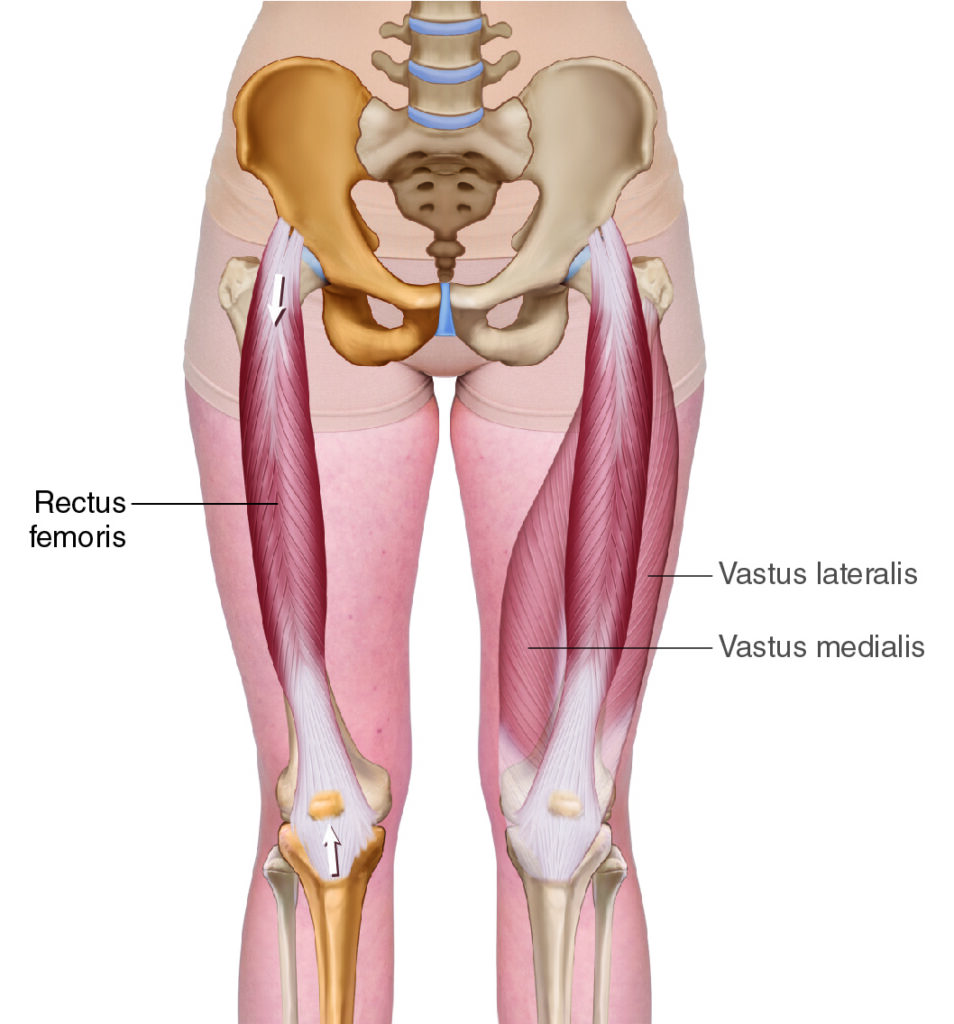
Rectus femoris. Permission Dr. Joe Muscolino (learnmuscles.com).
The rectus femoris is one of the four quadriceps femoris muscles, and the only one that crosses the hip joint as well as the knee joint. Whereas the other quad muscles run at an angle, the rectus femoris runs straight down the thigh from the pelvis to the tibia, so the word rectus is in the name. Rectus means straight; think “rectify the matter” as in “get it straight“!
Rectus Femoris Attachments
The rectus femoris runs from the pelvis (origin) to the tibia (insertion).
More specifically, it attaches from…
proximally: anterior inferior iliac spine (AIIS) of the ilium of the pelvis
to
distally: tibial tuberosity, via the patellar tendon
Rectus Femoris Functions
Open-Chain
The rectus femoris crosses the knee joint anteriorly with a vertical direction to its line of force, so it extends the knee joint, (as do all the quad muscles), or more specifically, open-chain, it extends the leg at the knee joint.
The rectus femoris also crosses the hip joint anteriorly with a vertical direction to its line of force, so it flexes the thigh at the hip joint.
Closed-Chain
With closed-chain kinematics, the distal attachment is fixed/stable, so the proximal attachment moves toward the distal one. At the knee joint, this means that the anterior surface of the thigh moves toward the anterior surface of the (lower) leg. So, the thigh extends at the knee joint. This movement is actually extremely functional and very common. Whenever you want to stand up from a seated position, you must move the thighs into extension toward the legs at the knee joints.
This is actually why the quads, if considered to be one muscle, would, by far, be the largest muscle in the human body. Why? Not to extend the leg at the knee joint, but rather to extend the thigh at the knee joint to get up from a seated position.
At the hip joint, closed-chain mechanics result in the proximal attachment of the rectus femoris, in other words, the pelvis, moving toward the distal attachment. So, this would bring the anterior surface of the pelvis toward the anterior surface of the thigh, resulting in anterior tilt of the pelvis at the hip joint.
Anterior pelvic tilt is extremely important functionally, because when the rectus femoris is tight, the pelvis is pulled posturally into excessive anterior tilt, which then causes the lumbar spine to have an excessive lordotic curve of extension; this postural distortion pattern is known as lower crossed syndrome. The excessive lumbar lordosis itself creates greater weight bearing on the posterior facet joints, which can cause low back pain, and is known as facet syndrome.
Notes:
- Extension of the thigh at the knee joint is the closed-chain/reverse action of the open-chain extension of the leg at the knee joint.
- Anterior tilt of the pelvis at the hip joint is the closed-chain/reverse action of the open-chain flexion of the thigh at the hip joint.
Nearby Anatomy
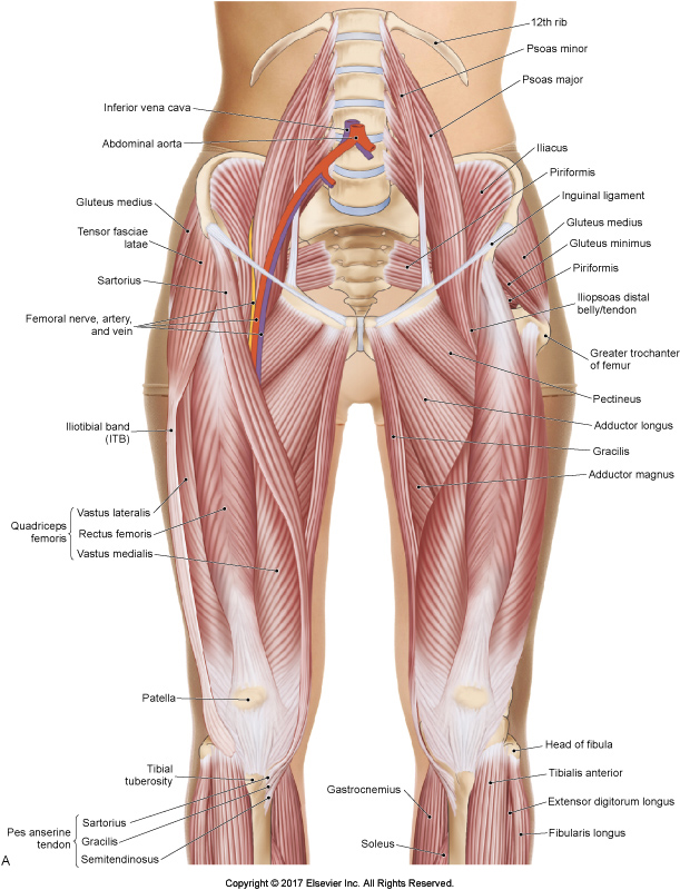
Permission Dr. Joe Muscolino – The Muscular System Manual 5ed.
The rectus femoris attachment onto the pelvis is between the tensor fasciae latae (TFL) and sartorius. The TFL lies lateral and the sartorius lies medial.
In the thigh, the vastus intermedius lies deep, the vastus lateralis lies lateral, and the sartorius and vastus medialis lie medial to the rectus femoris.
Note: All the quad muscles, but especially the rectus femoris, envelop the patella, and then attach into the tibial tuberosity via the patellar (also known as the infrapatellar) ligament. As such, this structure is considered to be both a tendon and a ligament.
Palpating the Rectus Femoris
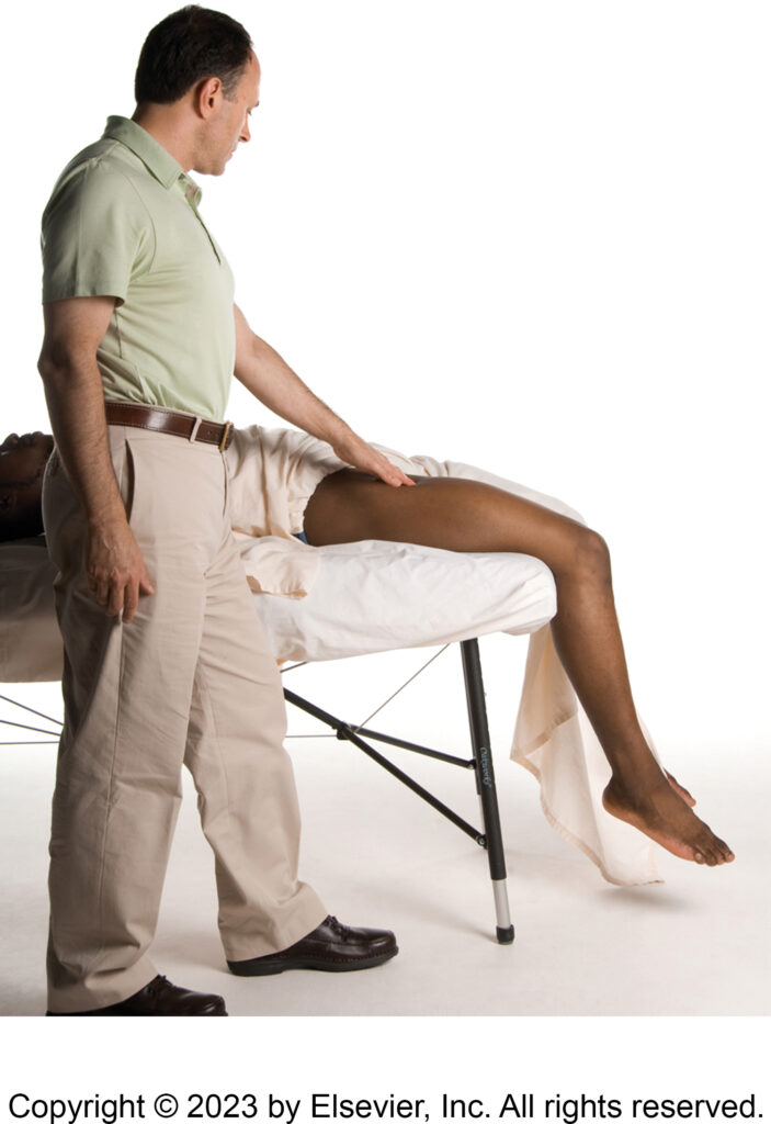
Optimal position for the lower extremity being palpated… thigh on the table, leg off the table.
Permission Dr. Joe Muscolino – The Muscle and Bone Palpation Manual 3ed.
The rectus femoris is superficial in the anterior thigh so is easy to locate, palpate and discern from adjacent musculature. With the client supine, start somewhere in the mid-thigh, dead center anteriorly, and you will be on the rectus femoris.
Ask the client to extend the (lower) leg at the knee joint and the rectus femoris (as well as the rest of the quadriceps) will engage and harden.
Follow it in baby steps (having the client contract and relax, feeling for engagement/hardening and relaxation/softening) all the way to the tibial tuberosity attachment.
Then return to your starting point and follow the same process all the way to the proximal AIIS attachment.
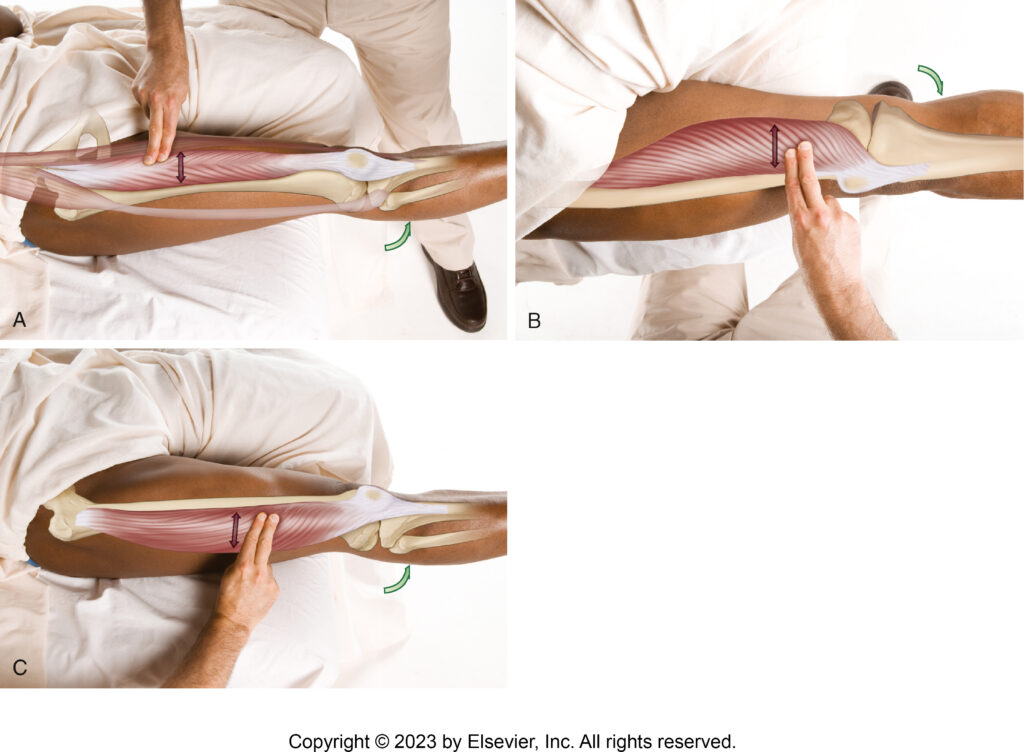
Palpation of the quads. Rectus femoris shown in figure A. Permission Dr. Joe Muscolino – The Muscle and Bone Palpation Manual 3ed.
The AIIS attachment can be a bit challenging to palpate and discern. This is usually best accomplished by holding the client’s thigh passively in flexion at the hip joint, allowing you to sink in deeper to the attachment on the AIIS.
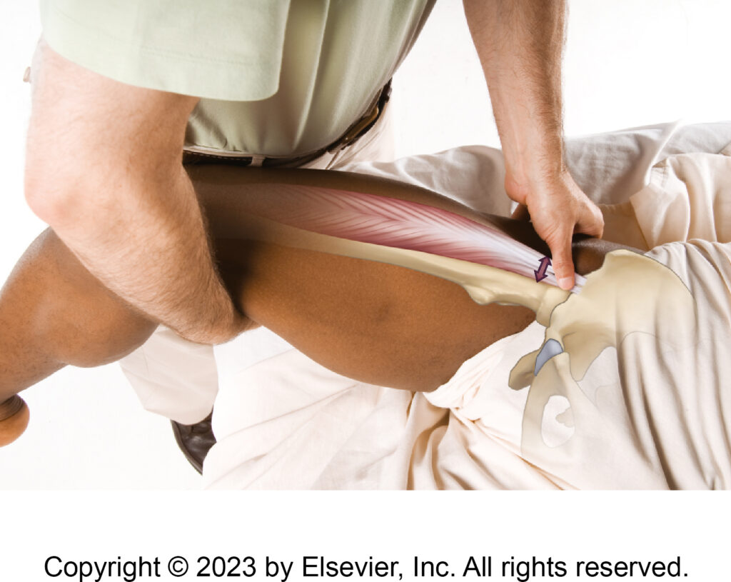
Palpating the AIIS attachment. Permission Dr. Joe Muscolino – The Muscle and Bone Palpation Manual 3ed.
Note: if the client is lying supine with their thigh on the table and their (lower) leg hanging off the table (as seen in the illustration), then it is easier to isolate knee joint extension from hip joint flexion. If this position is used, then it is important for the foot of the client’s other lower extremity to be placed on the table to help stabilize their pelvis and protect their low back (not seen in the illustration).
Important: This position should not be maintained for very long because it can be uncomfortable for the low back.
Stretching the Rectus Femoris
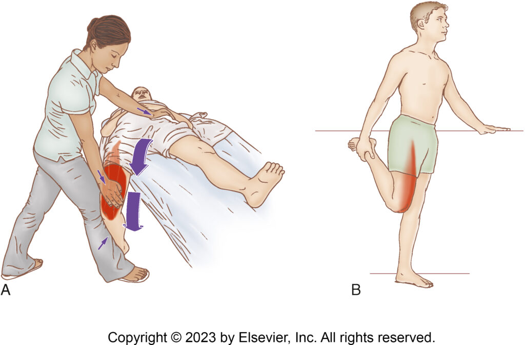
Stretching the rectus femoris. Permission Dr. Joe Muscolino – The Muscle and Bone Palpation Manual 3ed.
The rectus femoris flexes the hip joint and extends the knee joint, so it is stretched with full knee joint flexion accompanied by hip joint extension.
Massaging the Rectus Femoris
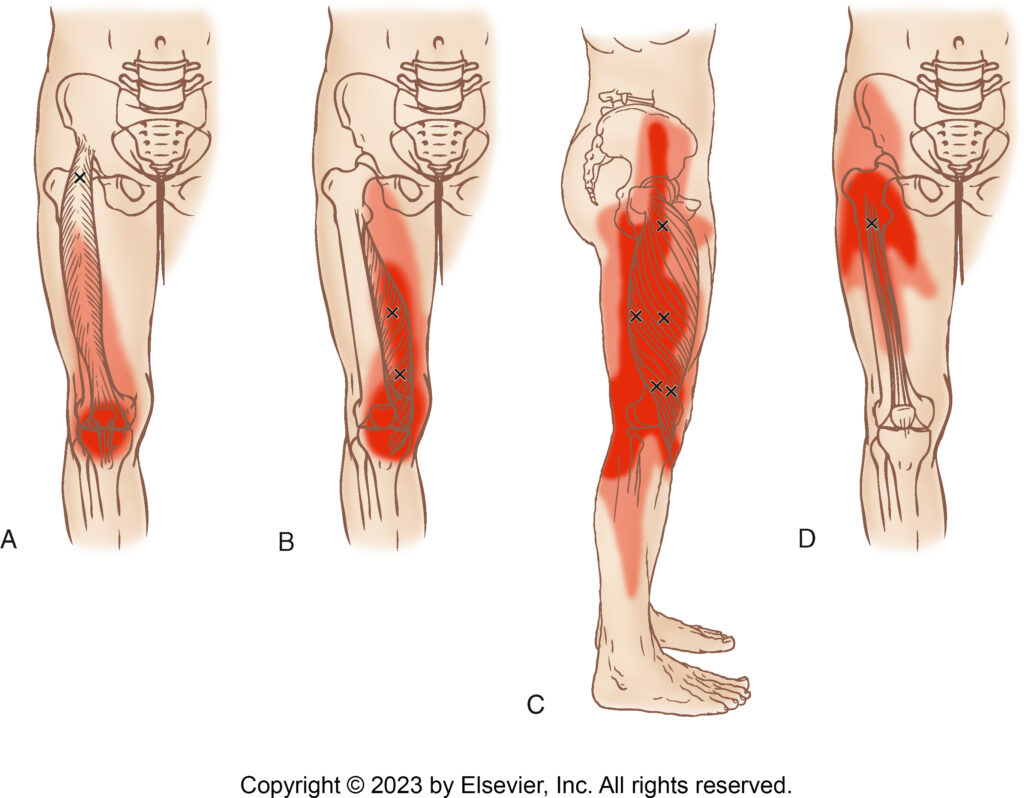
Quadriceps trigger points and their referral zones. Rectus femoris shown in figure A. Permission Dr. Joe Muscolino – The Muscle and Bone Palpation Manual 3ed.
Massaging the rectus femoris is easy to accomplish because so much of the muscle is superficial. It can be worked with any strokes you like: longitudinal (generally recommended to go from distal to proximal to follow the venous blood flow) or perpendicular. Deep friction perpendicular strokes through a towel are also very effective.
Because the AIIS attachment is a bit deep, it is usually best accessed with the client’s hip joint passively supported in flexion; placing a bolster under their knees can accomplish this nicely.
Conclusion
The rectus femoris runs straight up and down the anterior thigh, hence its name: rectus means straight. As a hip flexor, it is an extremely important muscle, both functionally and dysfunctionally. When tight, its pull is exerted in closed-chain function with anterior tilt of the pelvis; excessive anterior tilt can then create excessive lumbar lordosis (a postural distortion pattern known as lower crossed syndrome), which often predisposes toward low back pain.
Biography

Dr. Joseph Muscolino, DC is a soft-tissue oriented chiropractic physician and leading educator in manual and movement therapy. He is the author of eight major textbooks published by Elsevier and LWW, translated into more than 10 languages and used worldwide in core curriculum and clinical practice. A global lecturer and NCBTMB-approved CE provider, he offers COMT (Clinical Orthopedic Manual Therapy) certification workshops across the US and internationally. Visit his website at: LearnMuscles.com.
LearnMuscles Continuing Education (LMCE) is one of his online subscription platforms with over 4,000 video lessons for manual and movement therapy professionals, and more than 320 free NCBTMB-CE hours.
