
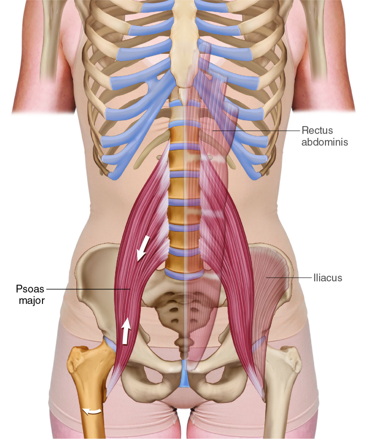
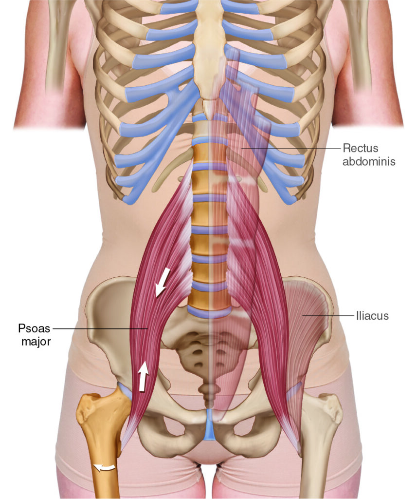
The Psoas Major. Permission Dr. Joe Muscolino (www.learnmuscles.com).
Quick Answer: The psoas major is a deep core/hip flexor muscle that runs from the anterolateral lumbar spine (T12–L5) to the lesser trochanter of the femur. It flexes and laterally rotates the thigh at the hip joint, and can flex, laterally flex, or (in extension) even extend the lumbar spine. This guide covers its attachments, joint actions, nearby anatomy, and step-by-step palpation, massage, and stretching protocols for manual therapists.
Table of Contents
- Introduction
- Psoas Major Attachments
- Psoas Major Functions
- Nearby Anatomy
- Palpating the Psoas Major
- Massaging the Psoas Major
- Stretching the Psoas Major
- Conclusion
- FAQ
Introduction
The psoas major is likely the most controversial and certainly one of the most important muscles in the human body. Running from the spine to the femur, it is functionally (and dysfunctionally) important for the lumbar spine and hip joints.
Psoas Major Attachments
The psoas major attaches from the anterolateral lumbar spine to the femur.
More specifically, the psoas major attaches…
anterolaterally on the bodies of T12 to L5, and the intervertebral discs between, and anteriorly on the transverse processes (TPs) of L1-L5
to the
lesser trochanter of the femur
Psoas Major Functions
The psoas major has many joint action functions, as well as stabilization functions. Following are concentric contraction/shortening joint action functions of the psoas major when the body is in anatomic position.
The psoas major crosses the hip joint anteriorly in the sagittal plane so it flexes the thigh at the hip joint (open-chain) and anteriorly tilts the pelvis at the hip joint (closed-chain).
The psoas major attaches onto the lesser trochanter in such a way that it has the ability to laterally rotate the thigh at the hip joint in the transverse plane (open-chain) and contralaterally rotate the pelvis at the hip joint (closed-chain). This is a minor joint action of the psoas major.
The psoas major crosses the lumbar spine anteriorly in the sagittal plane so it can flex the lumbar spine at the lumbar spinal joints (open-chain), and posteriorly tilt the pelvis at the lumbosacral joint in the sagittal plane (closed-chain).
The psoas major crosses the lumbar spine laterally in the frontal plane so it can laterally flex the lumbar spine at the lumbar spinal joints (open-chain), and elevate the same-side pelvis at the lumbosacral joint in the frontal plane (closed-chain).
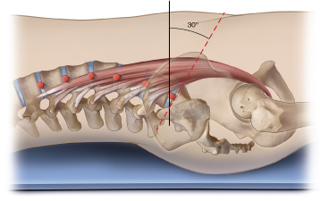
Line of pull of the psoas major for sagittal-plane motion – neutral position (sacral base angle of 30 degrees). – Permission Dr. Joe Muscolino (learnmuscles.com).
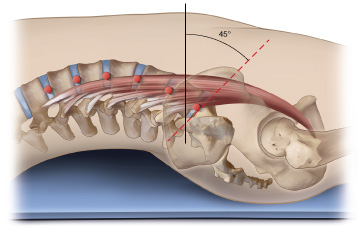
Line of pull of the psoas major for sagittal-plane motion – lumbar spine extended (sacral base angle of 45 degrees). Permission Dr. Joe Muscolino (learnmuscles.com).
Notes:
- It should be pointed out that the ability of the psoas major to flex the lumbar spine is based on the fact that (in anatomic position) its line of pull is anterior to the (mediolateral) mechanical axis for motion in the sagittal plane. But, if the lumbar spine is first in a sufficient amount of extension, the line of pull of the psoas major crosses posterior to the axis for sagittal plane motion, and therefore the psoas major would then actually extend the lumbar spine at the spinal joints. The lumbar spine might be in extension because the person happens to be moving into a position of extension at that moment, or because the person has a habitual posture of hyperlordosis of the lumbar spine, usually due to an excessively anteriorly tilted pelvis (essentially Vladimir Janda’s postural distortion pattern known as lower crossed syndrome).
- When in anatomic position (pelvic neutral – sacral base angle of approximately 30 degrees), the line of pull of the psoas major, although it is anterior to the axis for sagittal-plane motion, is only slightly anterior. Therefore, many sources state that it is actually more important for stabilizing the lumbar spine. But this stabilization also means that there is a good deal of compression placed upon the lumbar discs, therefore, the psoas major is often stated as contributing to intervertebral disc pathologies (e.g., bulging, herniation).
Nearby Anatomy
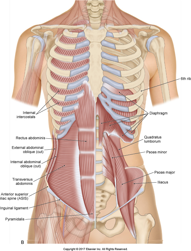
Musculature of the anterior trunk and thigh. Permission Dr. Joe Muscolino – The Muscular System Manual, 5ed.
The psoas major has its most superior attachment onto the T12 vertebra, so it passes through and interdigitates with the diaphragm.
The abdominal belly of the psoas major lies deep in the anterior abdominal cavity, directly in front of the lumbar spine. The psoas minor muscle (often absent) lies on the anterior surface of the psoas major (note: unlike the psoas major, the psoas minor does not cross the hip joint, so it has no function there).
The psoas major then crosses directly in front of the sacroiliac joint, contributing some attachment fibers into the ligamentous tissue of the anterior sacroiliac joint.
The femoral belly of the psoas major crosses the hip joint anteriorly, with the iliacus lying directly lateral to it, and the pectineus lying directly medial to it.
The femoral nerve, artery, and vein pass superficial to the femoral belly of the psoas major, so, caution should be exercised when working in this area.
The psoas major attaches on the lesser trochanter of the femur with the iliacus. For this reason, some sources describe these two muscles with the one name, the iliopsoas. If this terminology is used, it should be emphasized that although these two muscles have extremely similar function across the hip joint, the iliacus has no function at all at the spine, whereas the psoas major does. For this reason, my recommendation is to not group these two muscles together as the iliopsoas; rather, name them completely separately: the iliacus and the psoas major.
Palpating the Psoas Major
Palpation of the psoas major abdominal belly is challenging, but with experience, it becomes quite easy to perform with most clients. The easiest protocol to first learn is with the client supine, so that is what is presented here.
Abdominopelvic Belly:
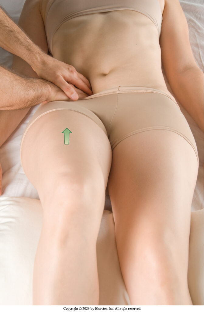
Supine palpation of the abdominopelvic belly of the psoas major. Permission Dr. Joe Muscolino – The Muscle and Bone Palpation Manual, 3ed.
Place a very large bolster under the client’s knees. This flexes their thighs at the hip joints, slackening their hip flexors, thereby allowing the pelvis to drop into posterior tilt, which slackens their anterior abdominal wall. Explain to the client that when you ask, they will need to raise their thigh into flexion a centimeter or so (a very small range of motion), to call upon the psoas major to engage.
Find the lateral border of the rectus abdominis and place your palpating finger pads directly lateral to it. Ask the client to take in a breath, and as they exhale and relax, sink in slowly, aiming for the anterolateral aspect of their spine. You are reaching for the psoas major against the spine. If necessary, you can reach the psoas in two or three excursions. When you believe you are there, check by asking the client to ever so slightly flex their thigh at the hip joint, and feel for the psoas major to engage and pop. Once located, move slowly in baby steps to locate the entirety of the muscle. Once located, you can assess the tissue health, feeling for any densifications (trigger points, fascial adhesions, etc.) in the muscle.
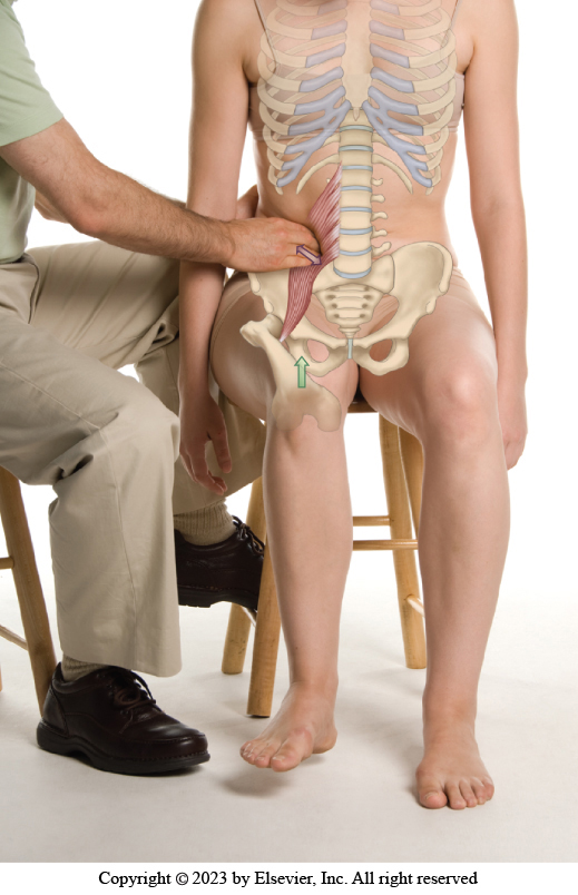
Seated palpation of the abdominal belly of the psoas major. Permission Dr. Joe Muscolino – The Muscle and Bone Palpation Manual, 3ed.
Notes:
- When palpating into the abdominal belly of the psoas major, for client comfort, it is extremely important to sink in SLOWLY, and work with the client’s breath.
- The psoas major abdominal belly can also be palpated with the client side-lying, seated, or even standing or prone, but the very best position is to have the client in a position in a ¾ side-lying, halfway between side-lying and supine. This allows you (with the table low) to use body weight to sink directly vertically downward into the belly.
Femoral Belly:
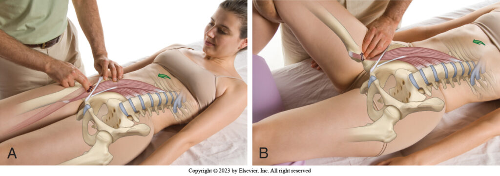
Supine palpation of the psoas major femoral belly and lesser trochanter attachment. Permission Dr. Joe Muscolino – The Muscle and Bone Palpation Manual, 3ed.
With the client supine and a bolster under their knees, find their ASIS and the pubic tubercle, and palpate approximately half way between along the inguinal ligament, feeling for the rounded/oval shape of the psoas major between the flatter iliacus on the lateral side and the flatter pectineus on the medial side. To confirm that you are on the psoas major, ask the client to do a gentle curl up, and the psoas major should engage. The key is that the psoas major is the only hip flexor that crosses the spine, and therefore the only hip flexor that will engage with spinal flexion (the curl up). Be sure to keep the curl up to be a very small excursion of movement.
Accessing the actual lesser trochanter attachment can be challenging, depending on the client. Best is to have their thigh flexed at the hip joint to slacken the hip flexor tissue. Then continue palpating the femoral belly in baby steps distally, with your pressure oriented medial-to-lateral toward the lesser trochanter.
Massaging the Psoas Major
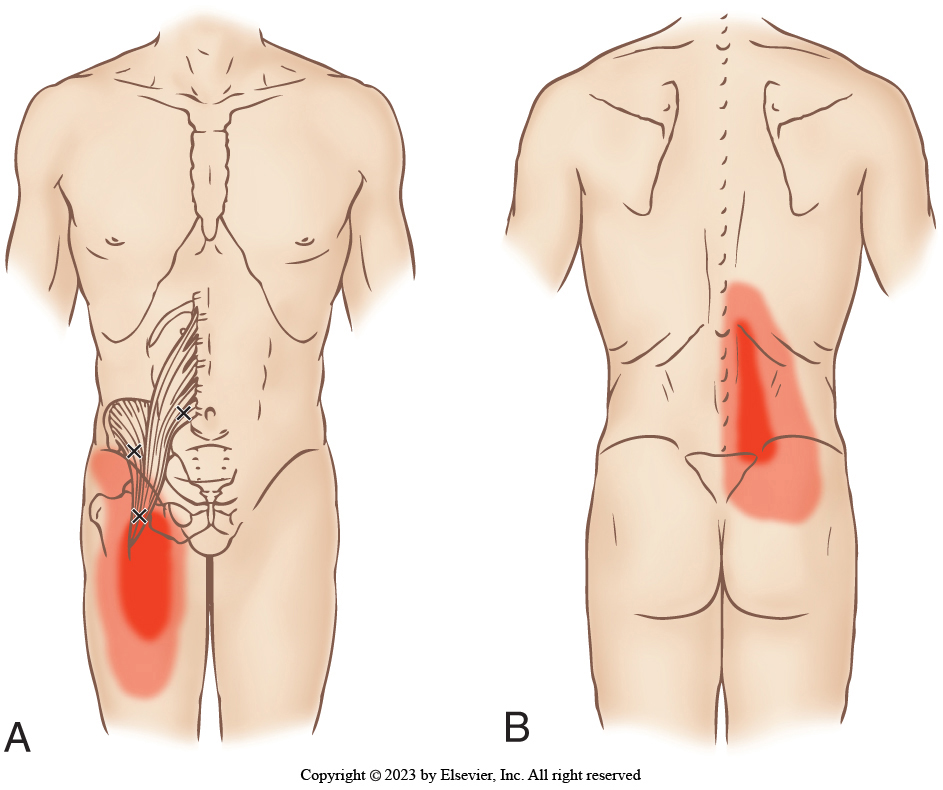
Psoas major trigger points and referral zones. Permission Dr. Joe Muscolino – The Muscle and Bone Palpation Manual, 3ed.
Massaging the psoas major, as will all muscles, follows from the palpation protocol. I like to palpate the psoas major abdominopelvic belly with small circular strokes. This way you can approach the muscle from all angles, to better assess and work it. Because this muscle is so deep, and very sensitive in many clients, it is extremely important to work slowly and with the client’s breath.
The femoral belly can be worked with strokes in any direction, from longitudinally along it, to perpendicularly across it, to any diagonal between.
Caution: The femoral nerve, artery, and vein lie superficial to the femoral belly of the psoas major, so approach this muscle slowly with your protocol, feeling for the pulse of the femoral artery, or any undue sensitivity on the part of the client due to pressure upon the nerve. If the artery or nerve is compressed, then simply move a few millimeters away from it and work from there, perhaps adjusting the angle of approach, depending on which aspect of the belly you are looking to access.
Stretching the Psoas Major
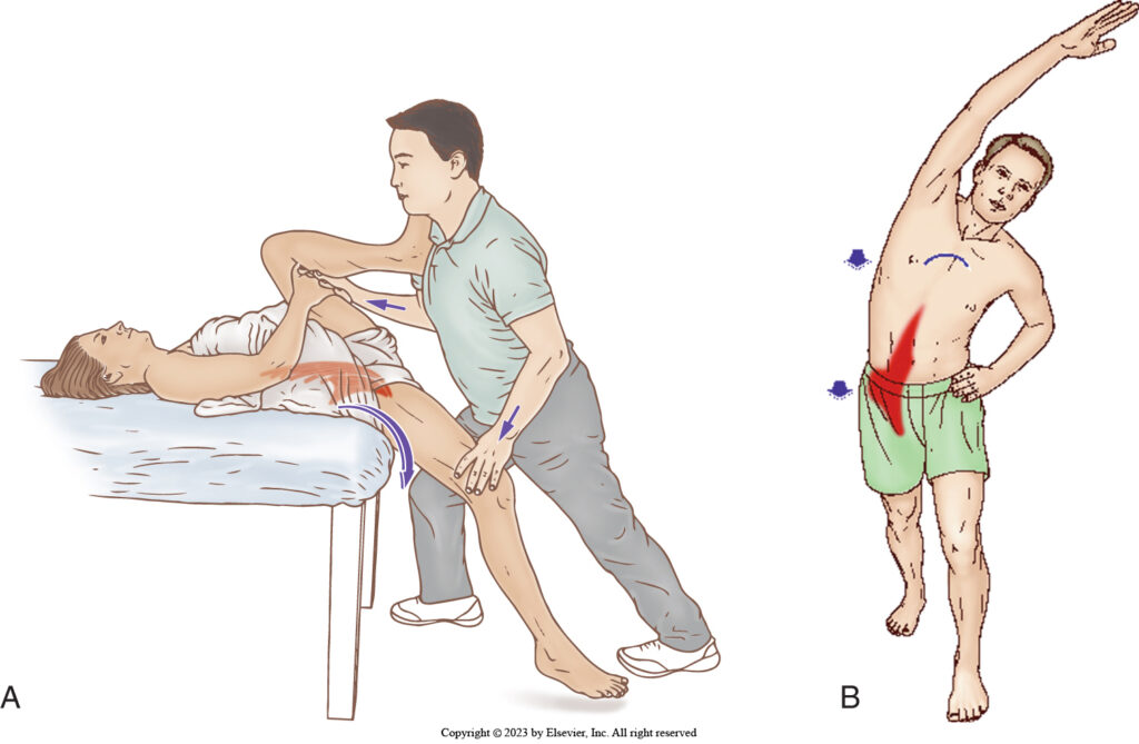
Therapist-assisted and client self-care stretching of the psoas major. Permission Dr. Joe Muscolino – The Muscle and Bone Palpation Manual, 3ed.
The psoas major can be best stretched by bringing the client’s thigh into extension. However, because this would theoretically stretch all of the many hip flexors, the stretch can be preferentially focused on the psoas major by having the client’s trunk/spine in lateral flexion to the opposite side.
Conclusion
Because the psoas major crosses the spine and hip joints, it is involved with pathomechanics of the spine and hip, making it one of the most important muscles in the human body to work with manual and movement therapy.
About the Author

Dr. Joe Muscolino (www.learnmuscles.com).
Dr. Joseph Muscolino, DC is a soft-tissue oriented chiropractic physician and leading educator in manual and movement therapy. He is the author of eight major textbooks published by Elsevier and LWW, translated into more than 10 languages and used worldwide in core curriculum and clinical practice. A global lecturer and NCBTMB-approved CE provider, he offers COMT (Clinical Orthopedic Manual Therapy) certification workshops across the US and internationally. Visit his website at: LearnMuscles.com.
LearnMuscles Continuing Education (LMCE) is one of his online subscription platforms with over 4,000 video lessons for manual and movement therapy professionals, and more than 320 free NCBTMB-CE hours.
FAQ
What does the psoas major muscle do?
The psoas major flexes the thigh at the hip joint and anteriorly tilts the pelvis (closed-chain). It also has a minor role laterally rotating the thigh, and it can flex or laterally flex the lumbar spine — or even extend it if the spine is already in extension.
Where does the psoas major attach?
The psoas major attaches anterolaterally on the bodies of T12–L5 and their intervertebral discs, and anteriorly on the transverse processes of L1–L5, running down to the lesser trochanter of the femur.
Is the psoas major the same as the iliopsoas?
Not exactly. The iliopsoas is a combined term for the psoas major and iliacus, since both insert on the lesser trochanter. However, the iliacus has no function at the spine, while the psoas major does — so many educators recommend naming them separately.
How do you palpate the psoas major?
With the client supine and a bolster under the knees, palpate lateral to the rectus abdominis and sink in slowly on the exhale toward the anterolateral spine, confirming placement by feeling the muscle engage during a small hip flexion movement.
How do you stretch the psoas major?
Bring the client’s thigh into hip extension, and to preferentially target the psoas major over other hip flexors, add lateral flexion of the trunk/spine to the opposite side.
Why is the psoas major considered important in manual therapy?
Because it crosses both the lumbar spine and hip joint, the psoas major is heavily involved in spinal and hip pathomechanics, including postural distortions like lower crossed syndrome and contributions to disc compression.