
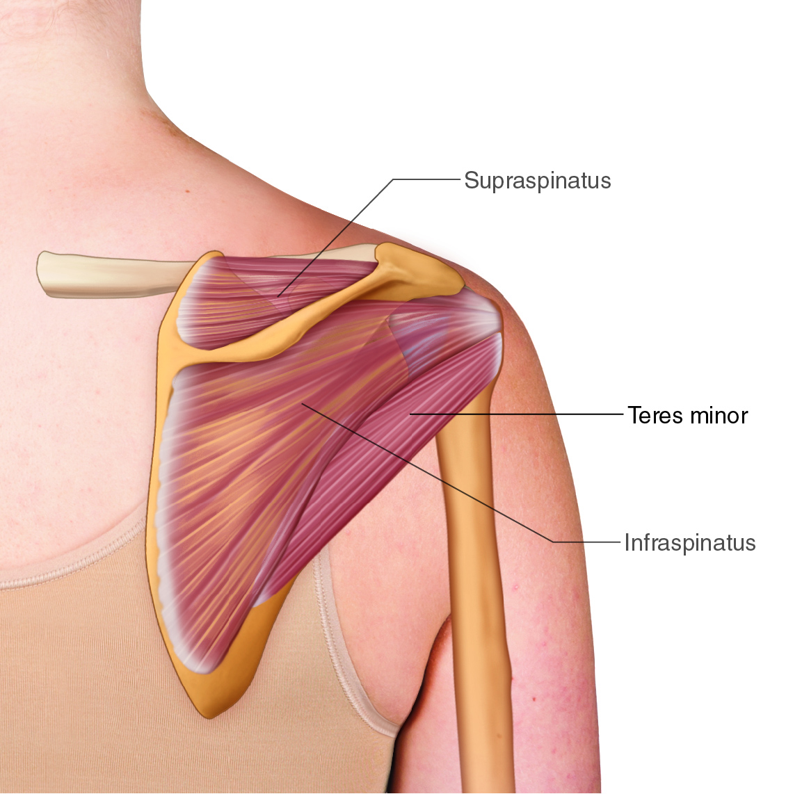
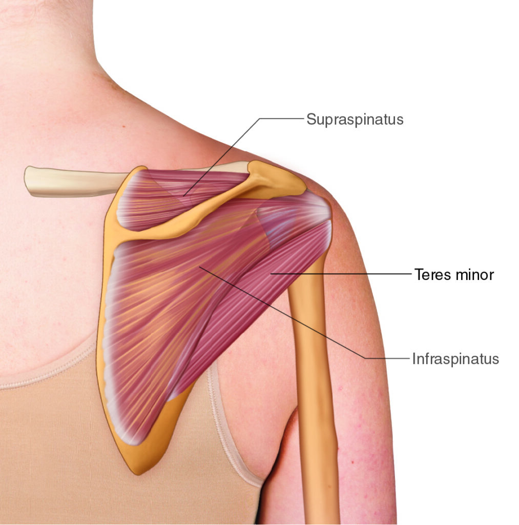
The Teres Minor. Permission Dr. Joe Muscolino (learnmuscles.com).
The teres minor is one of the four rotator cuff muscles of the shoulder (glenohumeral) joint, along with the infraspinatus, supraspinatus, and subscapularis. It attaches from the superior two-thirds of the lateral border of the scapula to the greater tubercle of the humerus. Its primary job is to laterally (externally) rotate the arm and to isometrically stabilize the humeral head in the glenoid fossa during shoulder movement. Because of this stabilizing workload, it is frequently involved in rotator cuff pathology and often has myofascial trigger points, making it an important muscle for manual and massage therapists to assess, palpate, and treat.
Table of Contents
- Introduction
- Teres Minor Attachments
- Teres Minor Functions
- Nearby Anatomy
- Palpating the Teres Minor
- Role in Rotator Cuff Pathology
- Massaging the Teres Minor
- Stretching the Teres Minor
- Conclusion
- FAQ
- About the Author
Introduction
The teres minor is one of the four rotator cuff muscles of the shoulder/glenohumeral (GH) joint. Due to the tremendous load placed upon this group for stabilization of the GH joint, it is often implicated in rotator cuff pathology.
The other three rotator cuff muscles are the infraspinatus, supraspinatus, and subscapularis.
Teres Minor Attachments
The teres minor attaches from the lateral scapula to the head of the humerus.
More specifically, it attaches from the:
superior 2/3 of the lateral border of the scapula
to the
greater tubercle of the head of the humerus
Teres Minor Functions
The teres minor has both a concentric/shortening function as well as an isometric stabilization function.
Concentrically, when the teres minor contracts and shortens, its joint action is to laterally (externally) rotates the arm/humerus at the GH joint. It shares this joint action with the nearby infraspinatus (another rotator cuff muscle).
And because the teres minor lies such that its line of pull is a bit inferior to the axis of motion of the GH joint in the frontal plane, it can also contribute to adduction of the arm at the GH joint.
When it contracts isometrically (staying the same length), it functions to stabilize the head of the humerus at the GH joint. This stabilization function is very interesting. From anatomic position, whether the humerus flexes, extends, abducts, or adducts, its distal end must lift. For this to occur, its proximal end, the head, must stay stabilized down into the glenoid fossa of the scapula.
If, for example, we picture the middle deltoid contracting to lift the humerus up into abduction (its distal end lifts up in the frontal plane), then we would see that the deltoid’s line of pull would also pull the proximal head straight up and it would jam into the acromion process above. To prevent this, and therefore for all of the deltoid’s pulling force to be focused on lifting the distal end of the humerus, we need to hold the head down into the glenoid fossa. The teres minor (along with the rotator cuff group as a whole) accomplishes this.
Therefore, the teres minor is isometrically active whenever the humerus lifts up into flexion and/or extension in the sagittal plane, abduction and/or adduction in the frontal plane, and concentrically active when the humerus is laterally rotating in the transverse plane (or adducting in the frontal plane).
For this reason, the teres minor (and indeed, all of the rotator cuff musculature) is often used/overused/misused/abused (to use the verbiage of educator Leon Chaitow), and therefore injured.
Nearby Anatomy
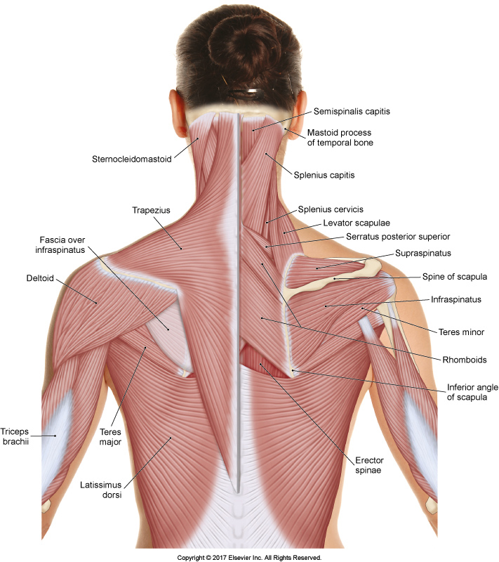
Permission Dr. Joe Muscolino – The Muscular System Manual, 5ed.
The belly of the teres minor usually blends into the belly of the infraspinatus, which lies directly inferior to the teres minor.
The teres minor lies directly superior to the teres major. Even though the teres major is next to the teres minor and shares the word “teres” in its name (“teres” is Latin for round), it is not related to it structurally or functionally. The teres major is not one of the rotator cuff muscles, and because it wraps around the humerus to attach in front, it medially rotates the humerus instead of laterally rotating it as the teres minor does.
On the greater tubercle of the humerus, the distal tendon of the infraspinatus attaches next to and superiorly to the teres minor. Superior to that, also attaching onto the greater tubercle, is the distal tendon attachment of the supraspinatus.
The posterior deltoid lies superficial to much of the teres minor.
Note: All four rotator cuff muscles attach onto the tubercles of the humerus. The supraspinatus, infraspinatus, and teres minor (SIT) onto the greater tubercle; and the subscapularis onto the lesser tubercle.
I often like to teach that if we know that all four rotator cuff muscles attach onto the two tubercles of the humerus, then we should be able to figure out that there must be three on the greater tubercle and one on the less tubercle. Why? Because if all four were to attach onto only one of the tubercles, there wouldn’t be the other tubercle (they are there as attachment sites for muscles). And if there were two on one and two on the other, then the tubercles would most likely both be the same size, so there wouldn’t be a greater and a lesser. So what choice is left? That there are three on one tubercle and one on the other; and three muscle must be on the greater tubercle because that is why it is greater (larger).
Palpating the Teres Minor
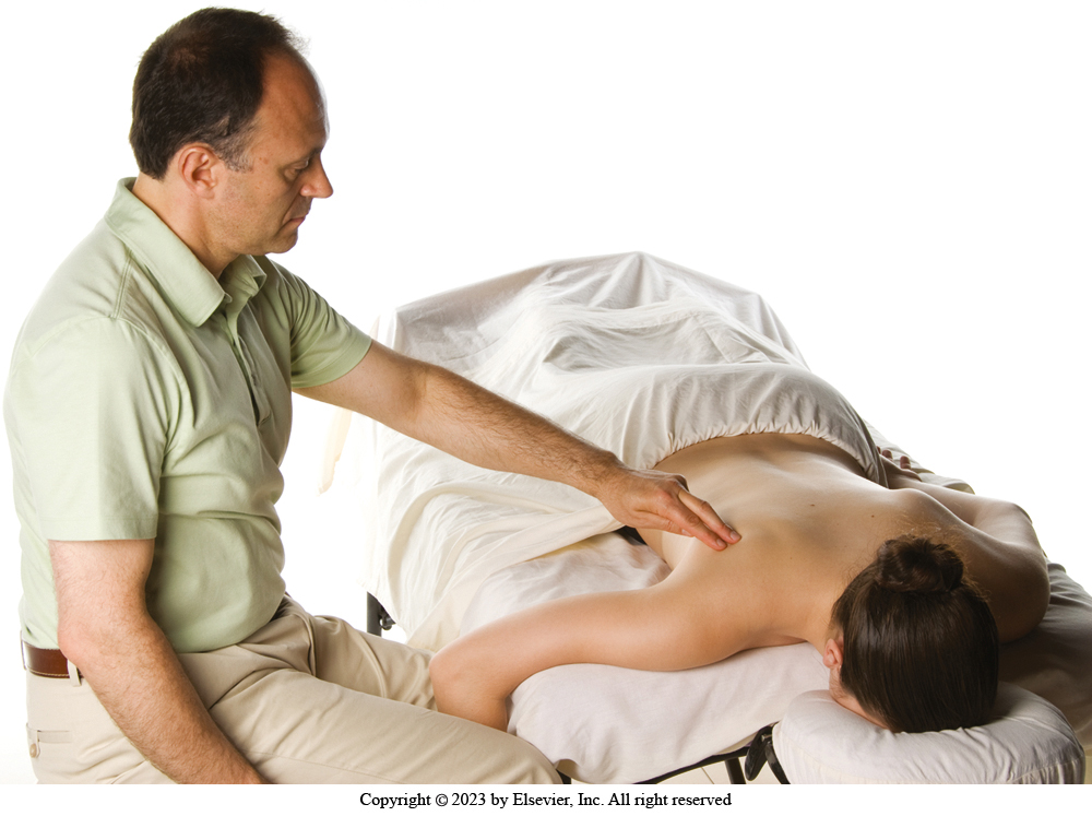
Palpating position for the teres minor. Permission Dr. Joe Muscolino – The Muscle and Bone Palpation Manual, 3ed.
To locate the teres minor, have the client prone on the table with their (upper) arm resting on the table and their forearm hanging off the table, with their distal forearm/hand between your knees. We will first locate the teres major, and then use that as a landmark to locate the teres minor.
Locate the inferior angle of the scapula, and come around to its lateral side. Now ask the client to medially rotate the arm at the GH joint by raising their forearm/hand toward your knee that is closer to the foot-end of the table. The teres major will engage/pop; strum perpendicular to it to confidently know its location.
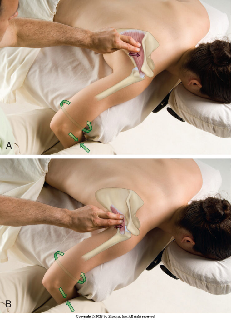
Palpation of the (infraspinatus and) teres minor. Permission Dr. Joe Muscolino – The Muscle and Bone Palpation Manual, 3ed.
Now palpate a bit more superiorly along the lateral border of the scapula and ask the client to laterally rotate their arm at the GH joint by raising their forearm/hand upward against your other knee (that is closer to the head-end of the table) (see illustration), and feel for the teres minor to engage/pop. Strum perpendicularly across the belly at the lateral scapula to strum (“twang”) it. Then move a baby step distally (laterally) toward its humeral attachment and repeat this protocol. Even though the distal tendon is deep to the posterior deltoid, it can usually be easily felt through the deltoid with a gentle lateral rotation engagement on the part of the client. Follow it as far toward the greater tubercle attachment as possible. Once the teres minor has been located, with it relaxed, palpate for density of the tissue.
Note: The same palpation protocol is used for the infraspinatus.
Role in Rotator Cuff Pathology
The teres minor is often implicated in rotator cuff pathology. Because the rotator cuff musculature is so intimately involved in GH joint stabilization, they are often (in the verbiage of Leon Chaitow) used/overused/misused/abused. In other words, they experience repetitive overuse syndrome. Rotator cuff pathology usually exhibits initially as tendinitis, but often progresses to degeneration, and then tearing. This can lead to instability of the GH joint, which can then result in muscle spasming as a protective splinting mechanism by the nervous system, which can then sometimes lead to frozen shoulder. Of the four rotator cuff muscles, the supraspinatus and infraspinatus are the two most commonly involved in rotator cuff pathology, but when the infraspinatus is involved, the teres minor is often involved as well.
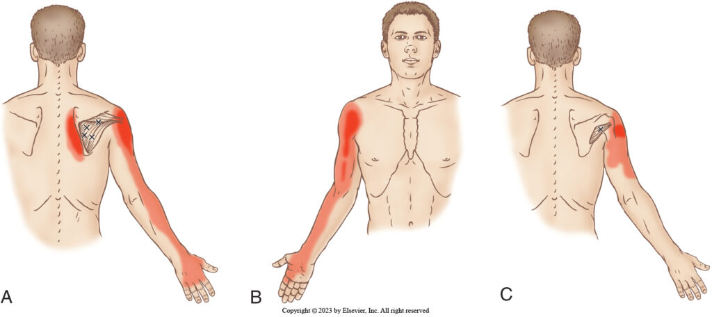
Infraspinatus and teres minor triggers points and their referral zones. Permission Dr. Joe Muscolino – The Muscle and Bone Palpation Manual, 3ed.
Even when the teres minor has not progressed to being part of a rotator cuff pathology, it very often contains myofascial trigger points that can cause pain for the client.
Massaging the Teres Minor
Massaging the teres minor follows from its palpation protocol. Once you have located it along its path from the scapula to the humerus, you can work it. There is no magic massage stroke that is always better for any one muscle or another, but cross fiber work is often performed across tendons. For the belly against the lateral scapular border, you can choose any stroke you would like; I very much like using circular strokes here.
The teres minor is often tight with myofascial trigger points, so your palpation assessment should be thorough and your massage and stretching should be performed effectively. Working these trigger points can often be tender for the client, especially if the client has never had the teres minor worked before. So, begin your treatment sessions for the teres minor with light to medium pressure, gradually increasing the depth of your work so that the client has time to allow for deeper pressure.
Stretching the Teres Minor
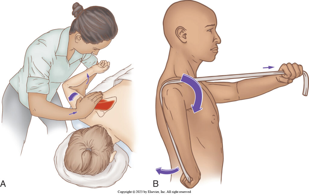
Stretching the infraspinatus and teres minor (therapist-assisted and client self-care). Permission Dr. Joe Muscolino – The Muscle and Bone Palpation Manual, 3ed.
Given that the teres minor is a lateral rotator of the arm at the GH joint, it can be stretched with medial rotation (a muscle’s joint action is its concentric/shortening function, and stretching is making the muscle longer, so simply doing the opposite of its joint action is how to stretch it). Given the challenge of medially rotating the humerus, the stretch protocol can be effectively performed using a rope or band (see illustration).
Caution: The teres minor is a fairly delicate muscle, so all the usual stretching precautions apply. Never stretch a muscle unless it has first been warmed up, never stretch it too far, and especially, never stretch a muscle fast. Warm up, stretch slowly and carefully, constantly feeling for the tissue response during the stretching protocol.
Conclusion
Given the tremendous load that is placed on the teres minor with everyday activities, and its role in rotator cuff pathology and how often it has myofascial trigger points, it is extremely important for the manual therapy practitioner to be competent and effective at working with this muscle.
FAQ
What is the teres minor muscle? The teres minor is one of the four rotator cuff muscles of the glenohumeral (shoulder) joint, alongside the infraspinatus, supraspinatus, and subscapularis. It stabilizes and laterally rotates the humerus.
Where does the teres minor attach? It attaches from the superior two-thirds of the lateral border of the scapula to the greater tubercle of the head of the humerus.
What is the main function of the teres minor? Concentrically, it laterally (externally) rotates the humerus at the glenohumeral joint. Isometrically, it stabilizes the head of the humerus in the glenoid fossa during other shoulder movements.
How do you palpate the teres minor? With the client prone, locate the teres major first via medial rotation of the arm, then palpate slightly superior along the lateral scapular border and ask the client to laterally rotate the arm to feel the teres minor engage.
How is the teres minor different from the teres major? Despite the similar name and proximity, they are unrelated in function: the teres minor is a rotator cuff muscle that laterally rotates the humerus, while the teres major is not part of the rotator cuff and medially rotates the humerus.
Why is the teres minor important in rotator cuff pathology? Because it shares heavy stabilization demands with the rest of the rotator cuff, it is prone to overuse, tendinitis, degeneration, tearing, and/or myofascial trigger points — especially when the nearby infraspinatus is also involved.
How do you stretch the teres minor? Since it’s a lateral rotator, it’s stretched via medial rotation of the humerus, often using a rope or band, always after warming up and performed slowly and carefully.
About the Author

Dr. Joe Muscolino (learnmuscles.com).
Dr. Joseph Muscolino, DC is a soft-tissue oriented chiropractic physician and leading educator in manual and movement therapy. He is the author of eight major textbooks published by Elsevier and LWW, translated into more than 10 languages and used worldwide in core curriculum and clinical practice. A global lecturer and NCBTMB-approved CE provider, he offers COMT (Clinical Orthopedic Manual Therapy) certification workshops across the US and internationally. Visit his website at: LearnMuscles.com.
LearnMuscles Continuing Education (LMCE) is one of his online subscription platforms with over 4,000 video lessons for manual and movement therapy professionals, and more than 320 free NCBTMB-CE hours.