A joint functions to allow motion, so two forms of joint dysfunction exist: Hypomobile joint has restricted motion; Hypermobile joint has excessive motion.
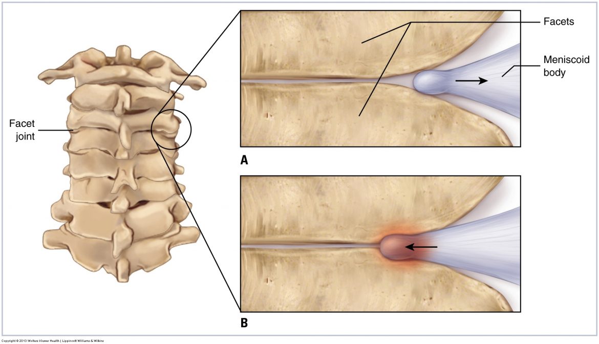
A joint functions to allow motion, so two forms of joint dysfunction exist: Hypomobile joint has restricted motion; Hypermobile joint has excessive motion.
When palpating a muscle and adding resistance to its contraction, never cross a joint that does not need to be crossed or other muscles will also contract.
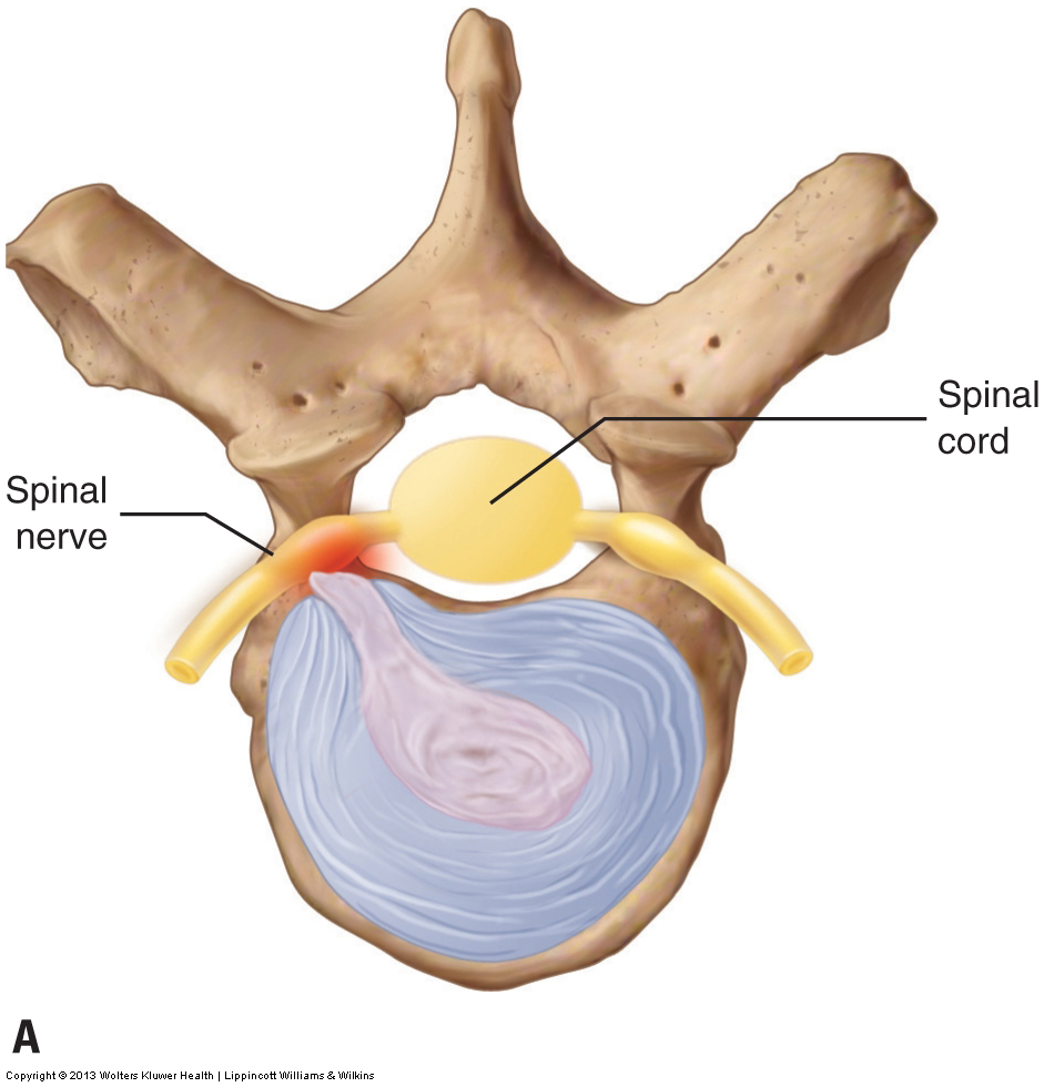
The danger with a disc bulge or herniation is that the disc can compress the spinal nerve within the intervertebral foramen, causing a pinched nerve.
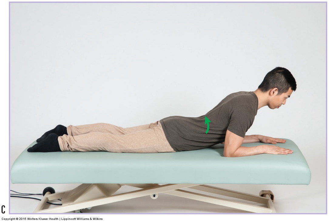
Self care for a herniated disc: avoid postures/activities that increase stress upon the disc and stretching/strengthening the musculature around the disc.
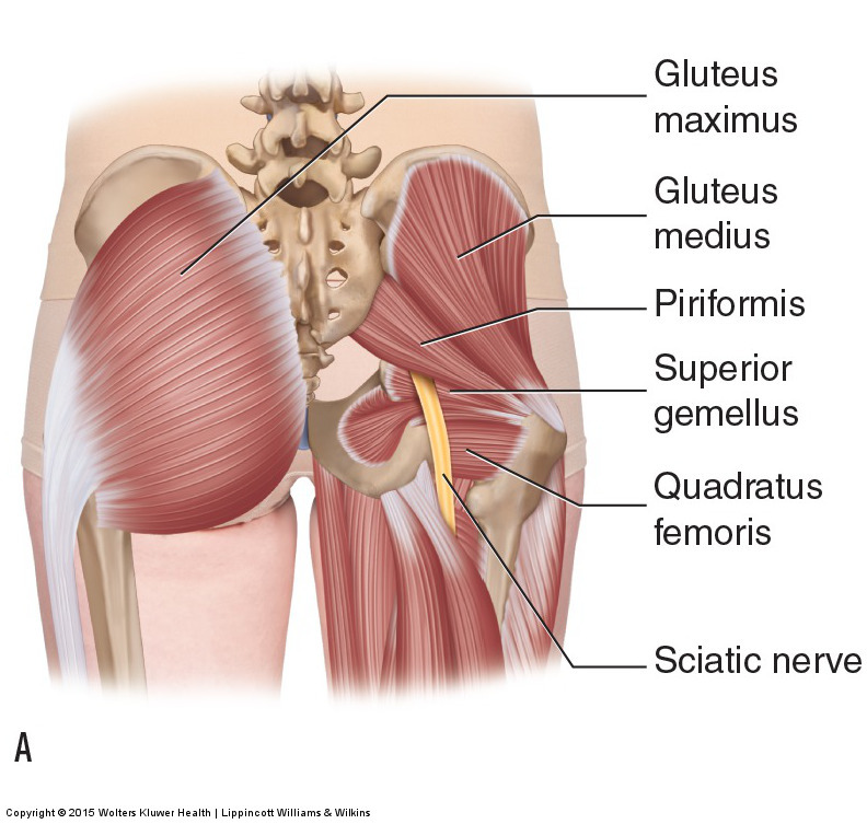
Piriformis syndrome occurs when a tight piriformis muscle compresses against the sciatic nerve, causing symptoms of sciatica into the lower extremity.
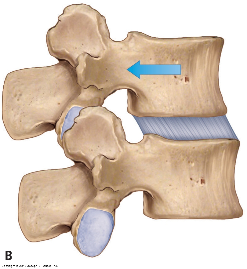
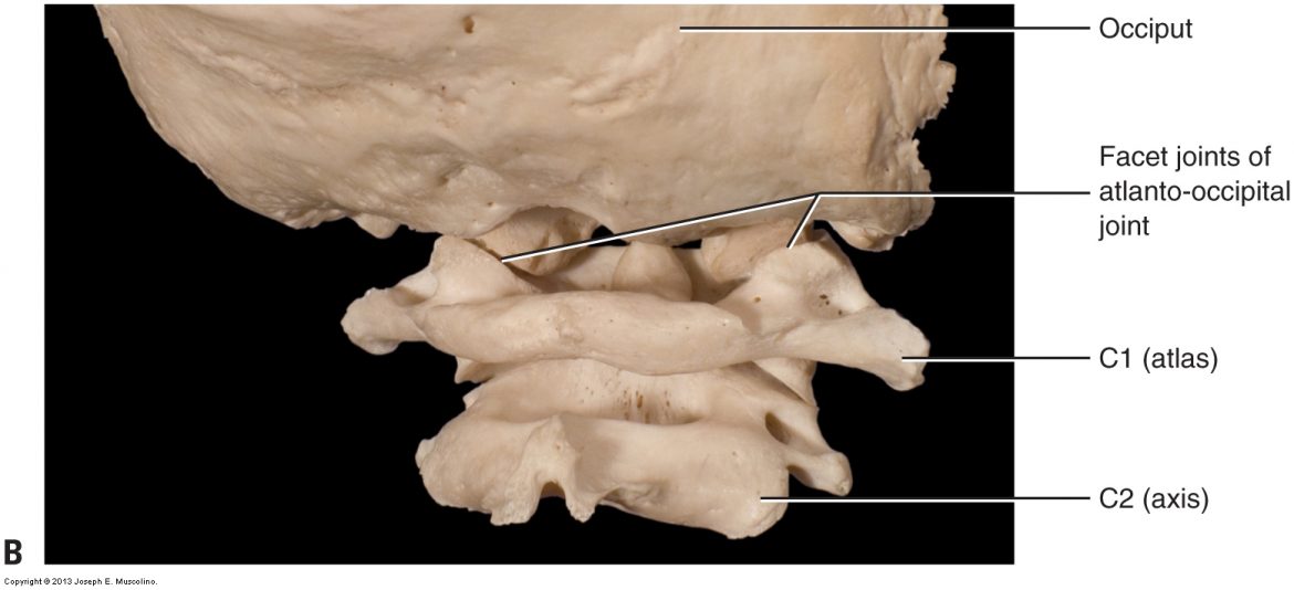
Table 1 shows average healthy ranges of axial motion of the cervical spine (head and neck), from the atlanto-occipital joint through the C7-T1 joint.
It is essential to exercise caution when working the anterior neck. However, this should not prevent therapeutic manual therapy work to the anterior neck.
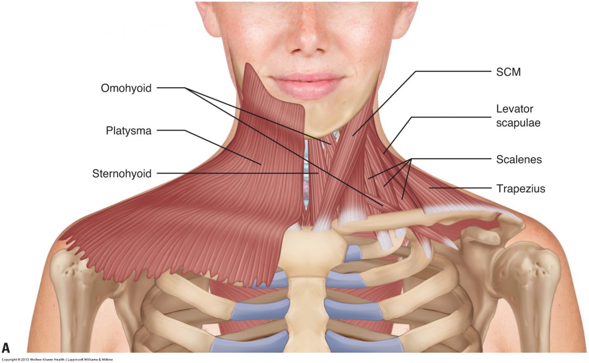
To perform orthopedic manual therapy to the neck that is accurate and specific, we need to know the attachments and actions of the muscles of the neck.
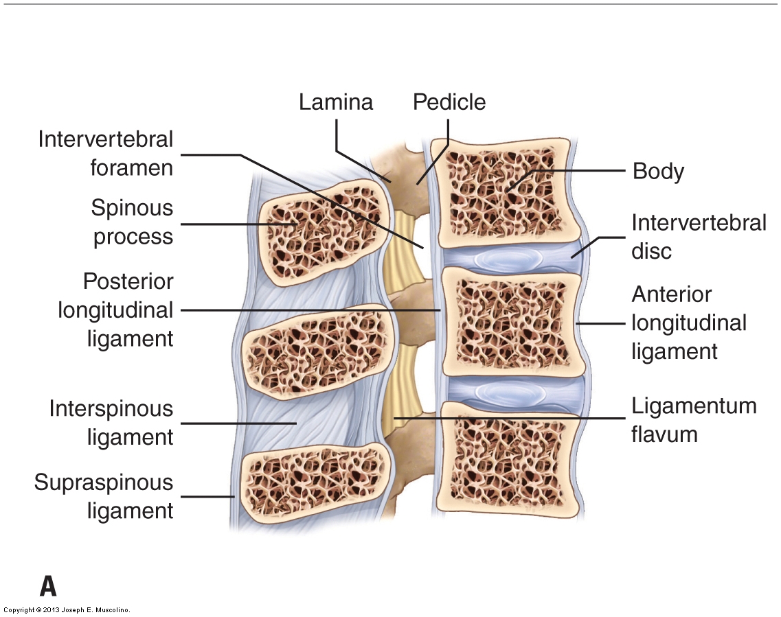
The “action” of a ligament is similar to that of an antagonist muscle. If either is tight/taut, it restricts motion in the opposite direction.
The disc joint is a cartilaginous joint that is composed of outer fibers called the annulus fibrosus that encircle the inner nucleus pulposus.
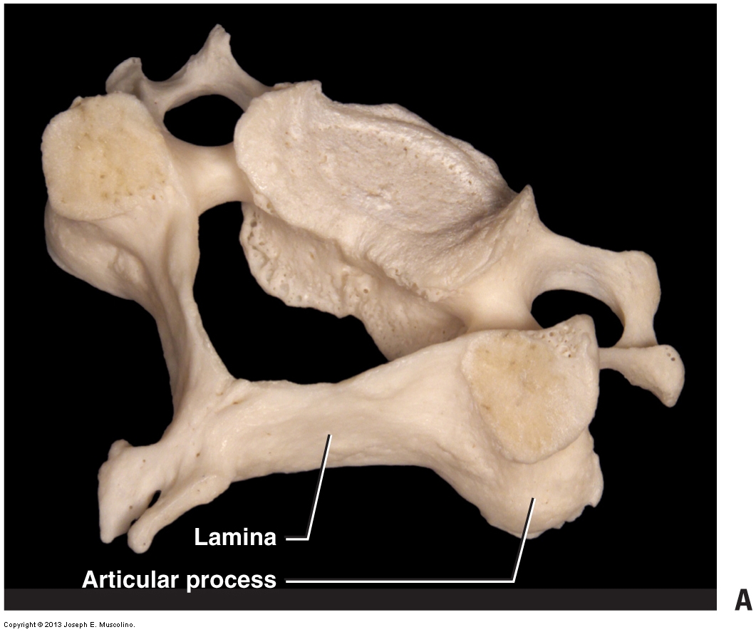
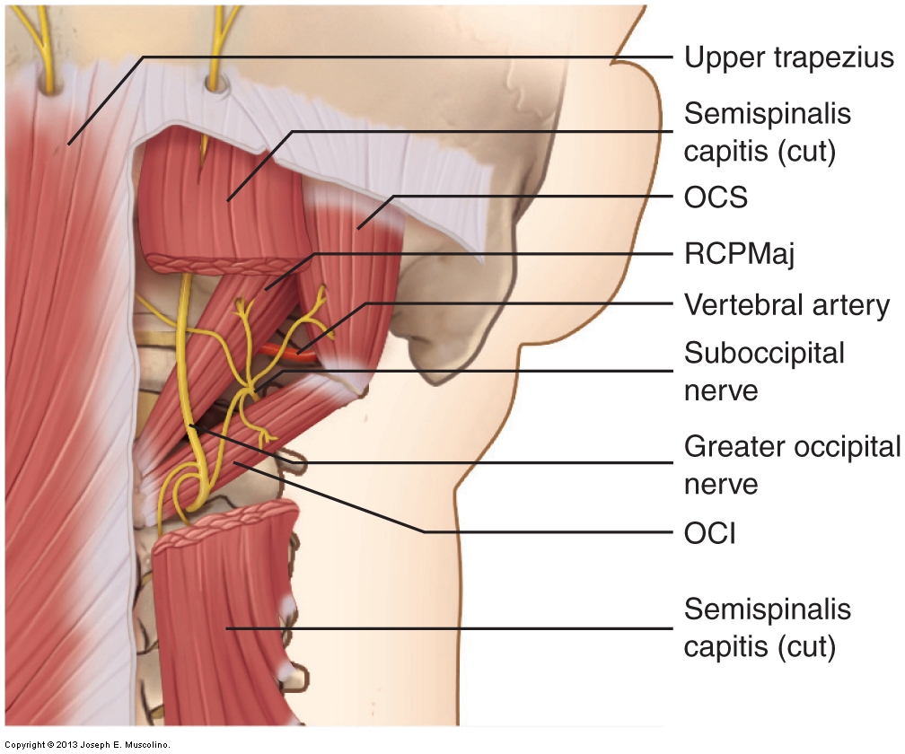
It is extremely important to be able to locate and palpate the laminar groove of the cervical spine because the thickest musculature is located there.
For manual therapy care to be successful, client communication is extremely important. This video explores who to effectively communicate with the client.
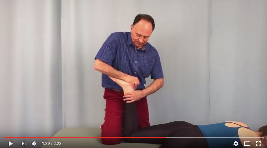
This video demonstrates Apley’s compression test for the knee joint, which assesses/diagnoses meniscus pathology.
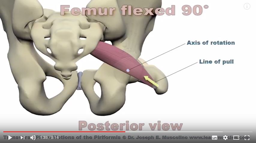
This 3D Animation demonstrates how the piriformis changes its joint action from being a lateral rotator of hip joint to being a medial rotator.
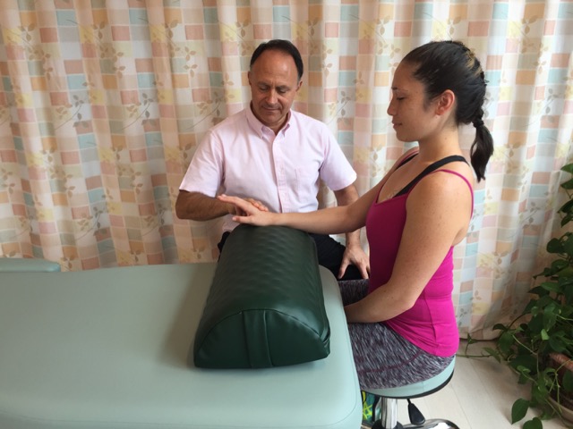
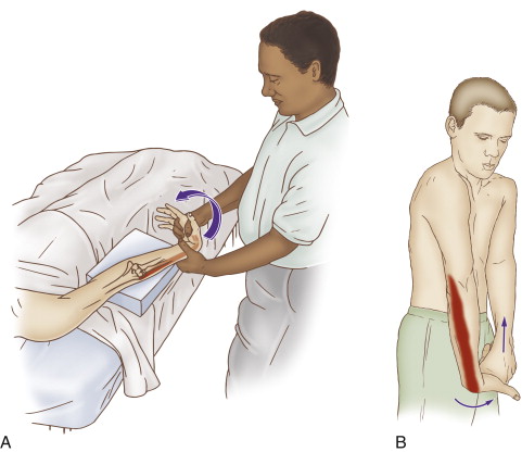
Assessment (diagnosis) of tennis elbow is done with active and passive range of motion, manual resistance, and palpation.
Self-care for tennis elbow should include frequent stretching of the hand and fingers into flexion. If inflammation is present, icing should be done.
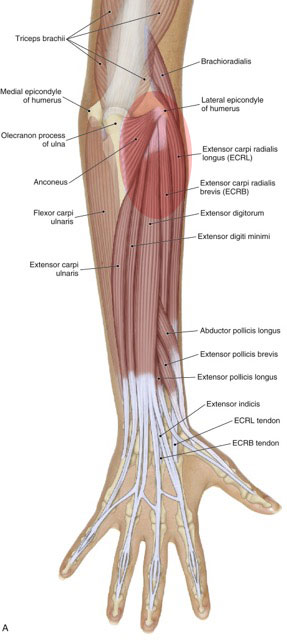
Symptoms of tennis elbow include pain and tightness at the common extensor belly/tendon, directly distal to the lateral epicondyle of the humerus.
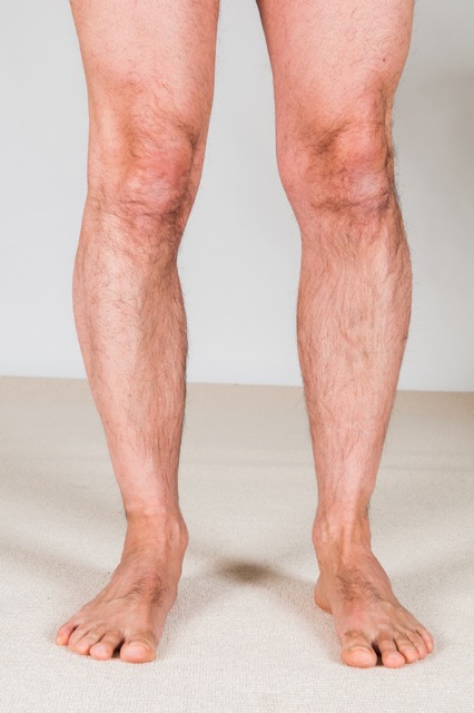
Case Study: Kerrati came in for wellness massage, but during the postural examination, the therapist noticed that her right arch drops markedly.
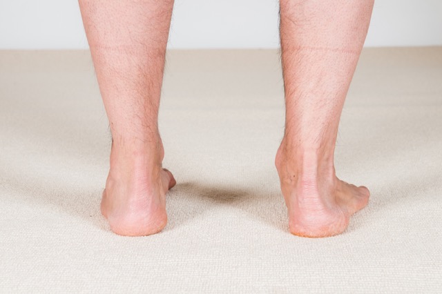
The first and most obvious sign of overpronation is a flat foot / dropped arch. A supple flat foot loses the arch only when weight bearing.