A healthy joint is both mobile and stable. However, some sources are concerned about the psoas major’s compression/stabilization effect upon the spine.
Below you'll find a list of all posts that have been categorized as “Uncategorized”
A healthy joint is both mobile and stable. However, some sources are concerned about the psoas major’s compression/stabilization effect upon the spine.
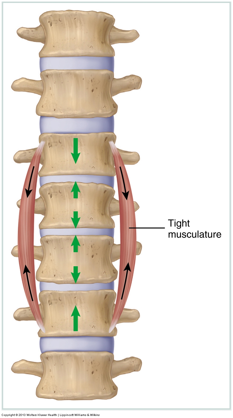
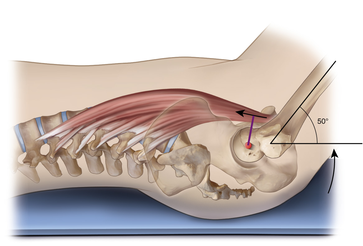
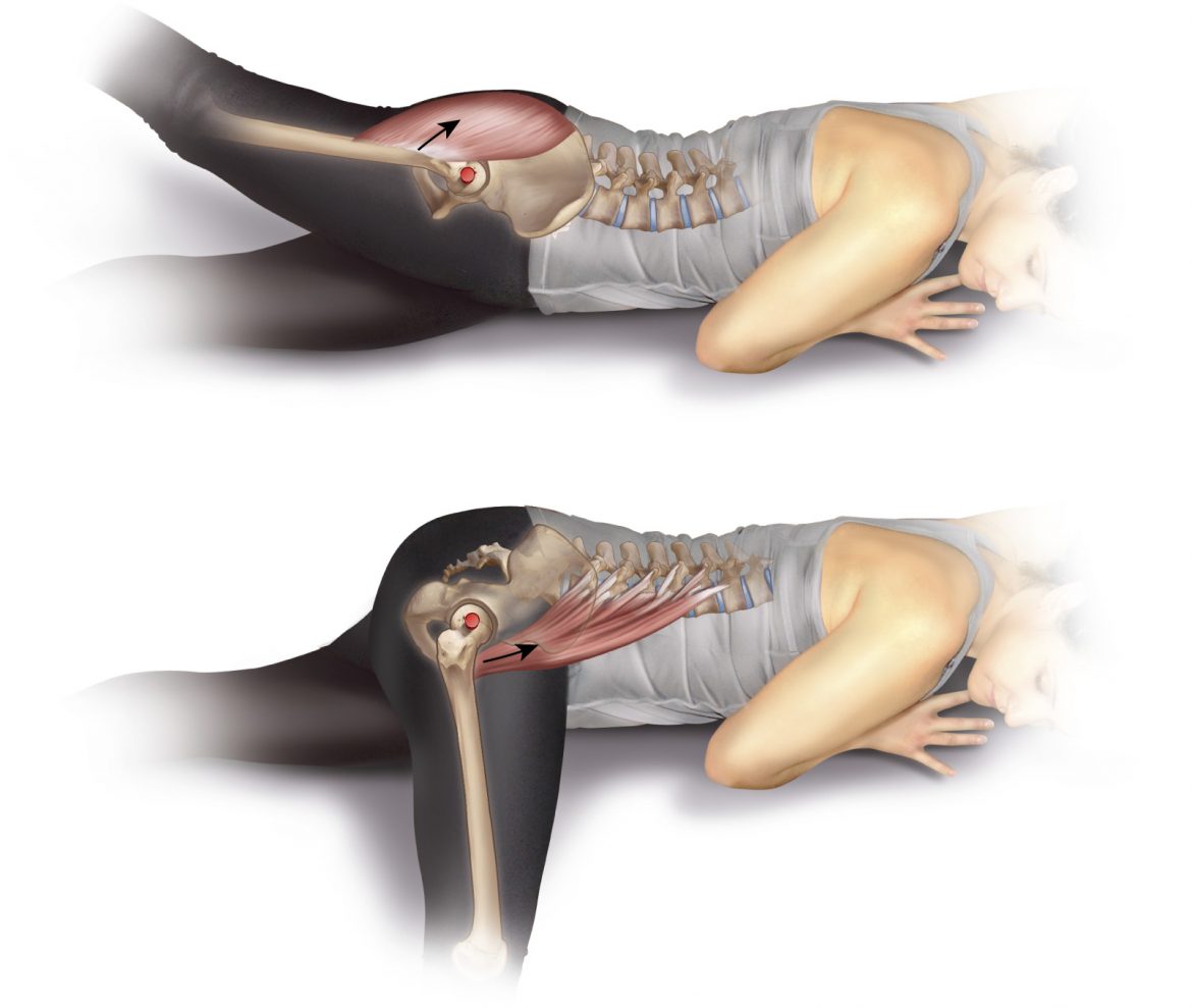
The psoas major’s sagittal plane lumbar spine joint action is by far its most controversial function. The term “psoas paradox” describes this controversy.
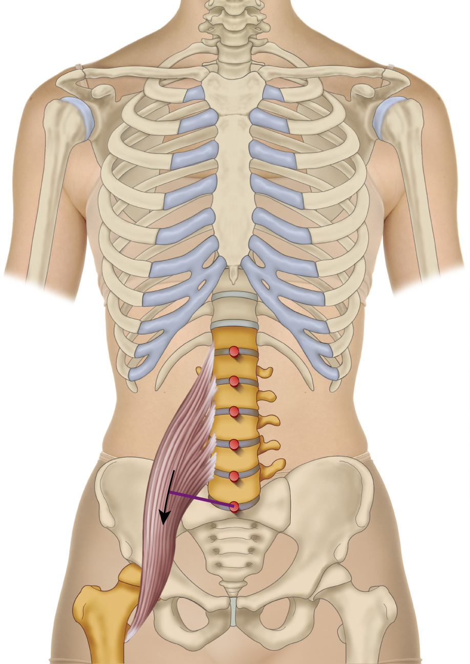
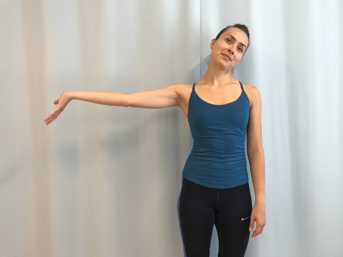
The frontal plane spinal action of the psoas major is fairly clear; it crosses the spinal joints laterally, so it lateral flexes the spine to that side.
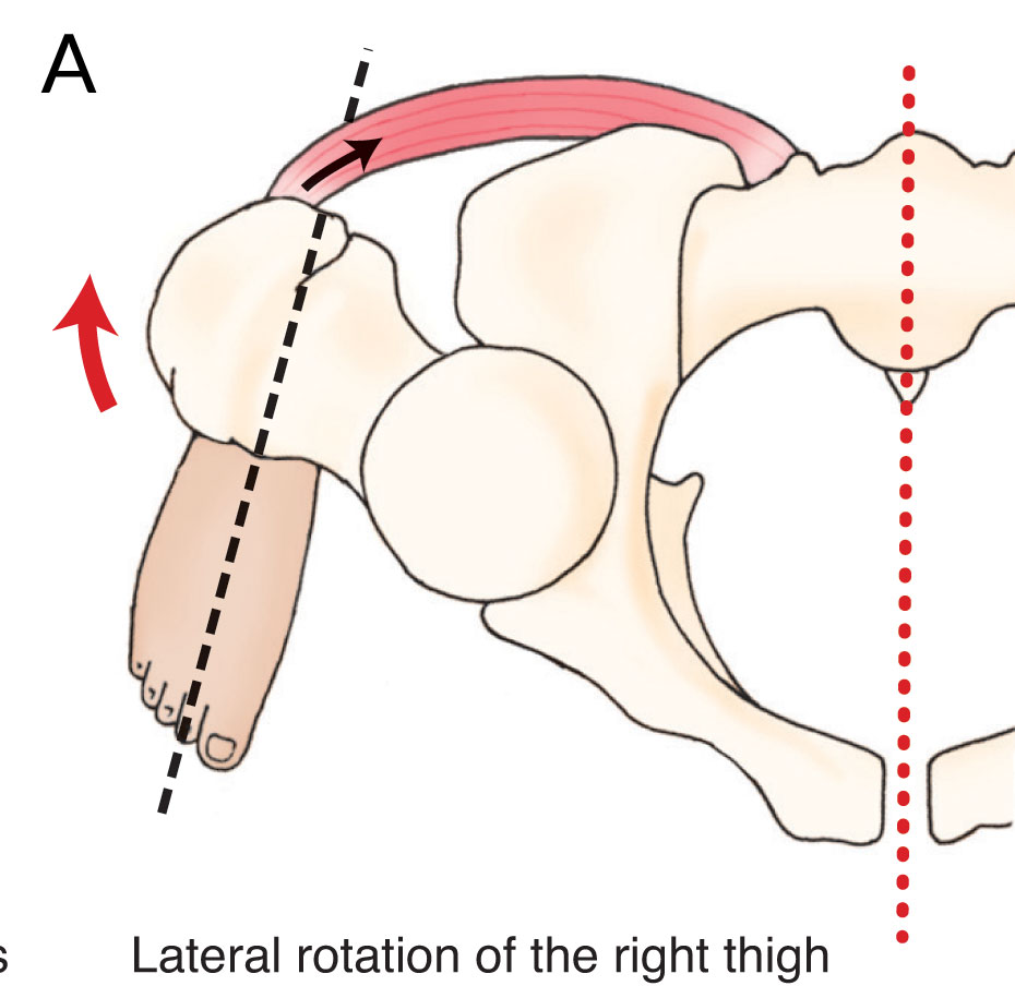
The function of the psoas major has been claimed to be both medial rotation and lateral rotation. However, most sources agree that it is a lateral rotator.
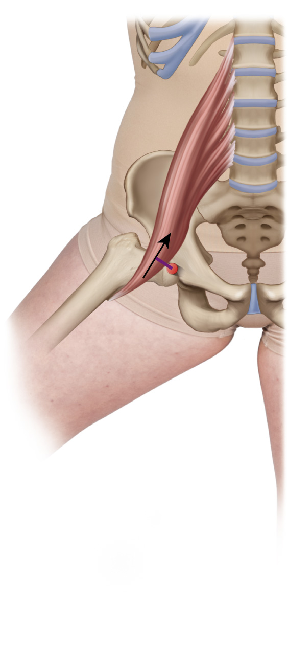
If the psoas major frontal plane open-chain action is abduction of the thigh at the hip joint, the closed-chain action is depression of the pelvis.
Note: This is the third blog post article in a series of 11 articles on Psoas Major Function. See below for the other articles in this series on psoas major function. The hip joint is a triaxial joint that allows …
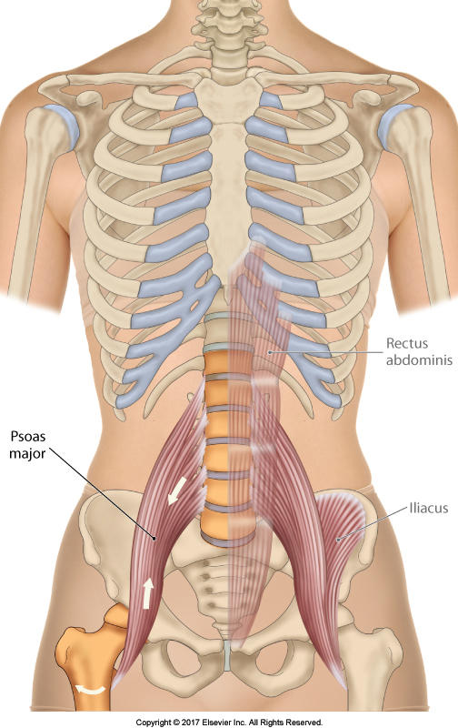
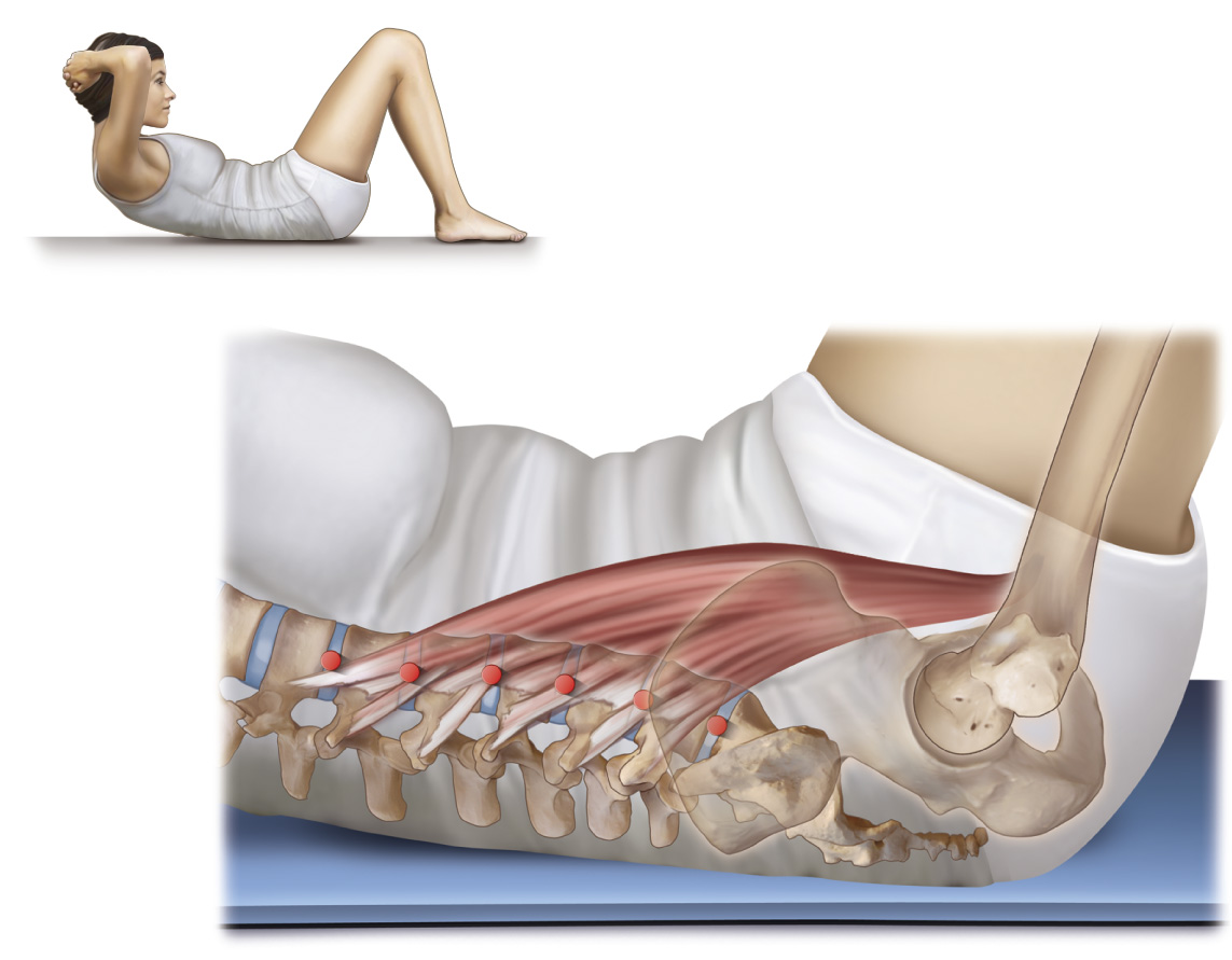
The psoas major is first and foremost, a muscle of the hip joint; however it is more complicated because it also crosses the spinal joints.
The Psoas Major may hold the distinction of being the most important as well as the most misunderstood muscle in the human body.
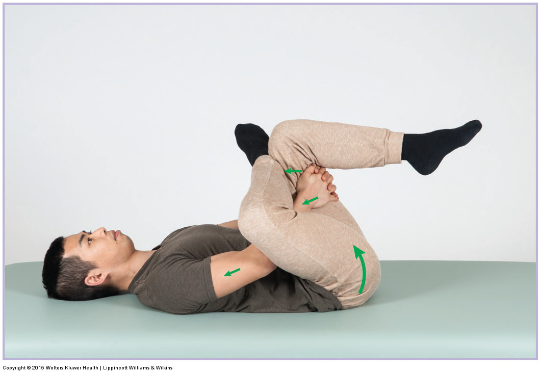
Self-care for piriformis syndrome involves heat followed by stretching. The client can perform either the Figure-4 or the horizontal adduction stretch.
This blog post introduces and explains how the manual therapist can perform assessment and treatment for specific musculoskeletal conditions of the neck.
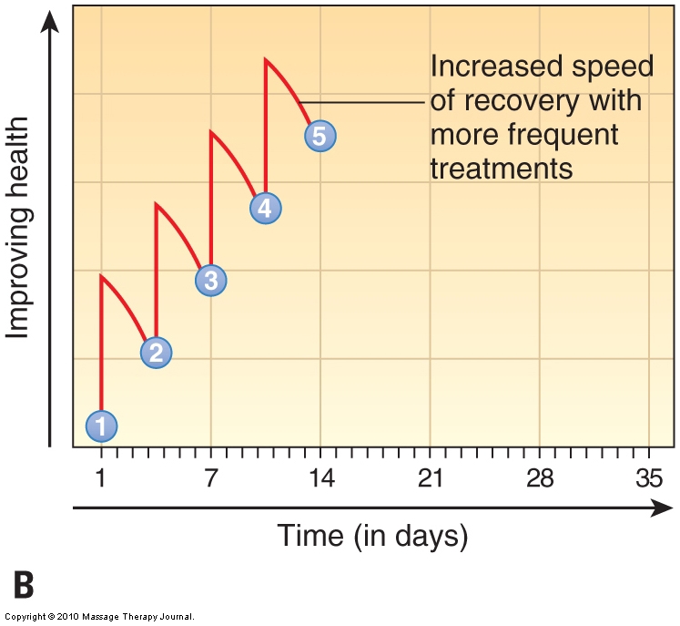
Sound Treatment Strategy: To perform effective manual therapy, frequency of care should be structured as in every other world of rehab: 2-3 x per week.
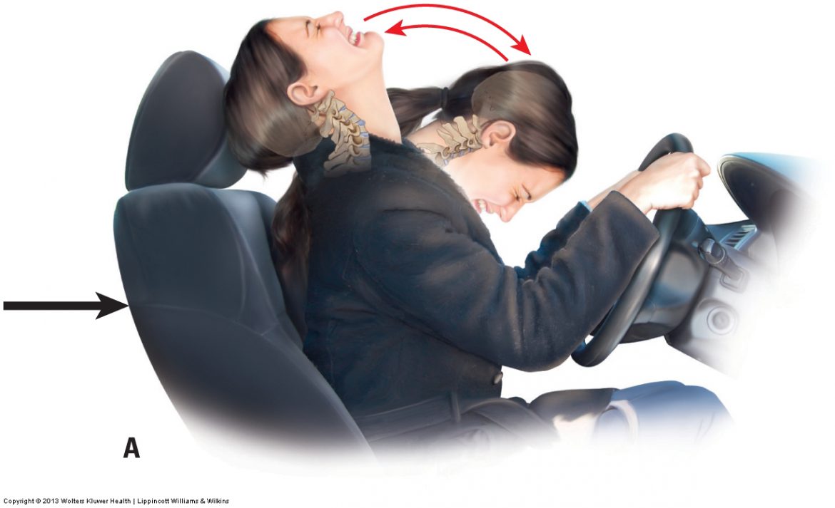
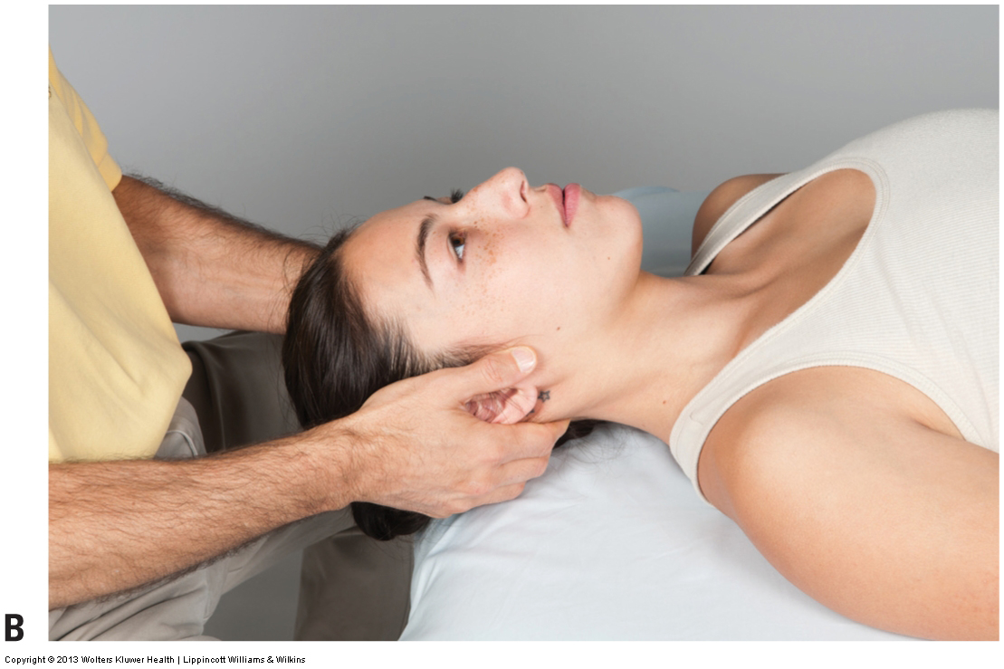
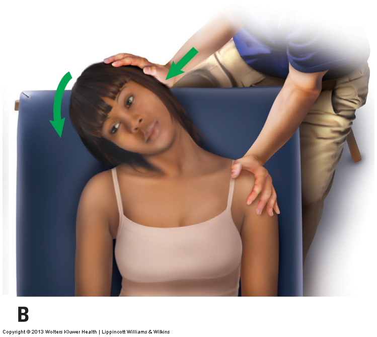
Vertebral artery competency test assessment is critically important to be performed if the neck will be stretched or mobilized during treatment.
The brachial plexus tension test (BPTT) is actually a series of three tests, each one designed to assess one of the three major nerves that enter the hand.
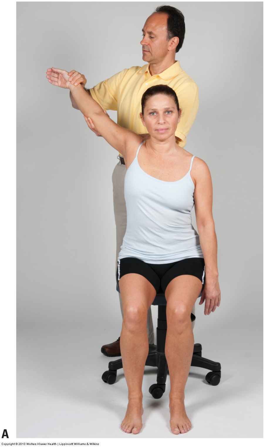
Thoracic Outlet Syndrome (TOS) is a very common set of posture dysfunctional patterns. Adson’s, Eden’s, & Wright’s tests are designed to assess TOS.
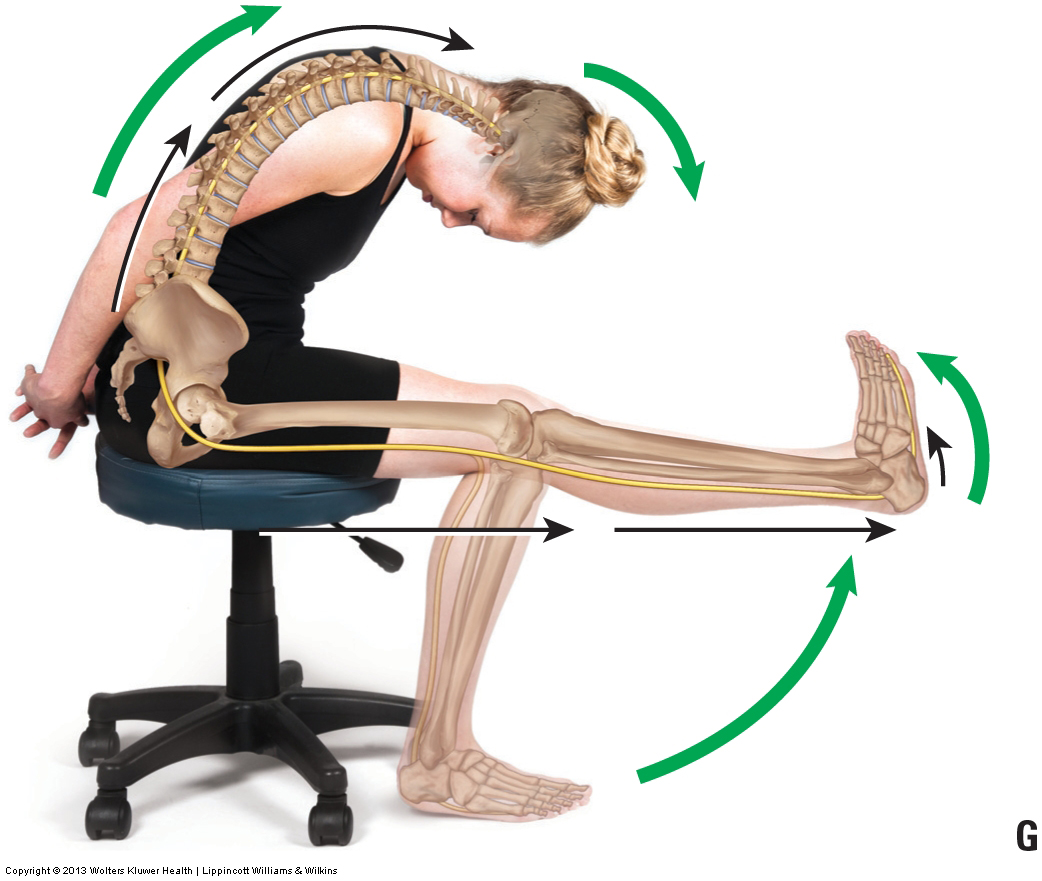
The slump test places tension on the spinal cord and PNS. It assesses a space occupying condition of the cervical and lumbar spine, as well as TOS.
The term “space occupying condition” is used to describe a condition in which there is compression on a spinal nerve in the intervertebral foramen.

Motion palpation is a specific form of passive (pin and stretch technique) assessment that challenges the intrinsic fascial tissue of the joint.

Perhaps no assessment procedure is more important to the manual therapist and integral to musculoskeletal (myofascioskeletal) assessment than palpation.
General orthopedic assessment testing includes three assessment techniques, two types of range of motion (ROM) assessment and manual resistance assessment.
Postural assessment: Good posture is balanced and symmetrical & does not place excessive stress on the body. Bad posture is asymmetrical and/or imbalanced.
If a physical examination assessment test creates signs/symptoms of the condition, it is positive and the therapist knows that tissue is unhealthy.
The health history should be done before the physical assessment exam because it helps reveal the regions that need to be assessed during the physical exam.
Before treatment can be given, it is necessary to have a clear understanding of the mechanism behind the musculoskeletal pathologic condition.
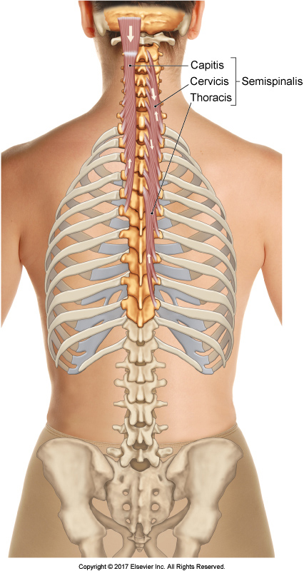
Semispinalis capitis, pectineus, & vastus lateralis are unusual suspect muscles, often overlooked by manual therapists, that can cause pain and dysfunction.
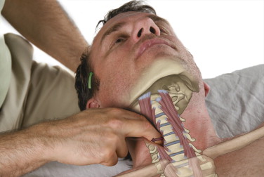
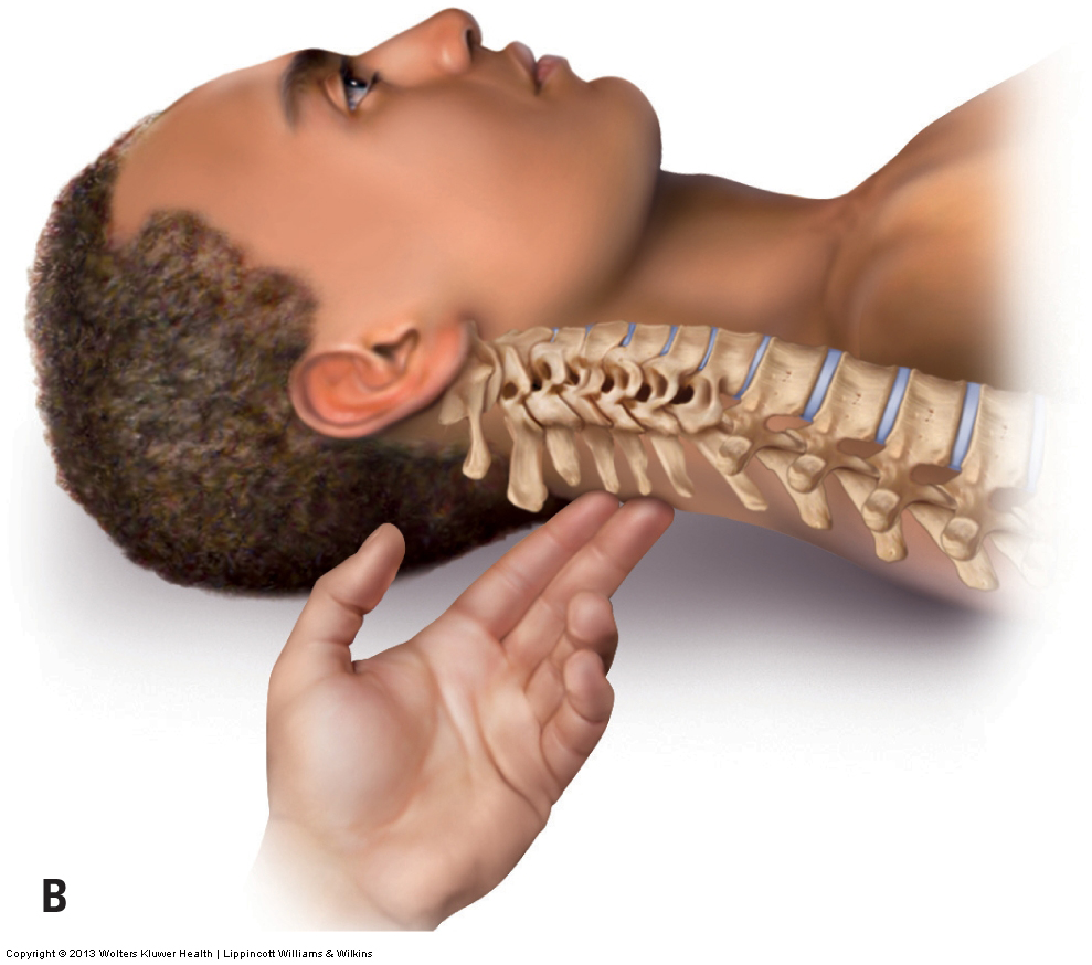
The longus colli and longus capitis are deep flexors of the neck that often become locked short and cause neck pain, especially when swallowing.
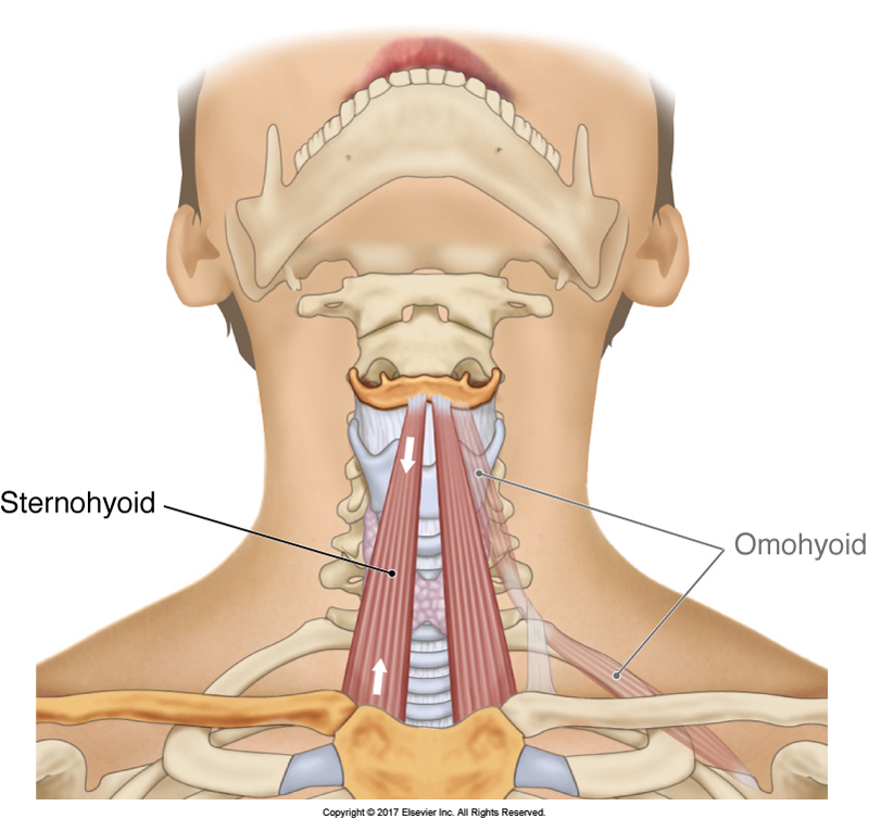
The sternohyoid is an infrahyoid muscle of the anterior neck that attaches from the sternum inferiorly to the hyoid bone superiorly and may cause neck pain.
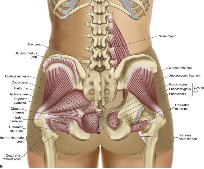
Attachments and Actions of the Coccygeus and Levator Ani The coccygeus and levator ani are pelvic floor muscles located between the sacrum and coccyx medially and the pelvic bone laterally (Figure 11). As pelvic floor muscles, they are important toward …
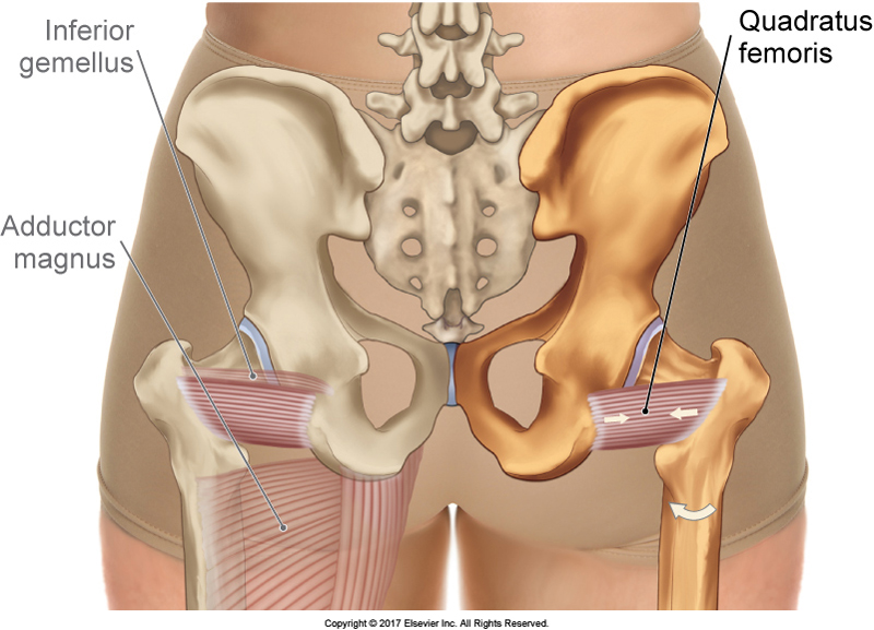
The quadratus femoris is a deep lateral rotator of the hip joint that is overlooked because of our excessive focus on its neighbor, the piriformis.
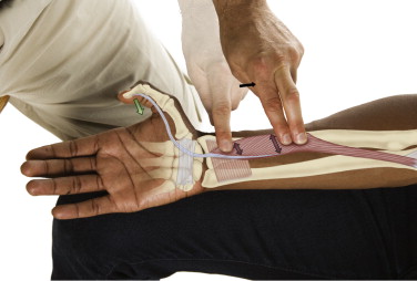
The flexor pollicis longus is a deep muscle of the anterior forearm and hand that flexes the thumb and is often overused with digital devices.
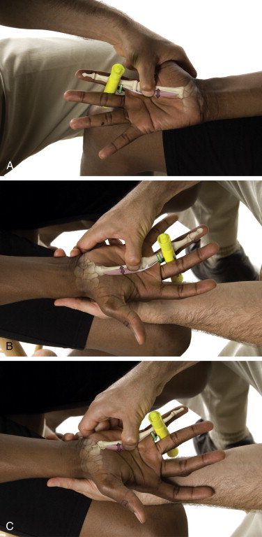
The palmar interossei are intrinsic hand muscles located between metacarpals bones in the palm of the hand. They may be the cause of hand pain.